👉 Click the link below and request access—I’ll approve it for you shortly!
https://www.notion.so/MSKMRI-KNEE-b6cbb1e1bc4741b681ecf6a40159a531?pvs=4
==============================================
✨ Join the channel to enjoy the benefits! 🚀
https://www.youtube.com/channel/UC4bw7o0l2rhxn1GJZGDmT9w/join
==============================================
👉 "Click the link to purchase on Amazon 🎉📚"
[Visualizing MSK Radiology: A Practical Guide to Radiology Mastery]
https://www.amazon.com/dp/B0DJGMHMFS
==============================================
MSK MRI Jee Eun Lee
📚 Visualizing MSK Radiology: A Practical Guide to Radiology Mastery Now! 🌟 Available on Amazon, eBay, and Rain Collectibles! 💻 Ebook coming soon – stay tuned! ⏳ 🔗 https://www.amazon.com/dp/B0DJGMHMFS 🔗 https://www.ebay.com/itm/3875004193
www.youtube.com
Visualizing MSK Radiology: A Practical Guide to Radiology Mastery
www.amazon.com

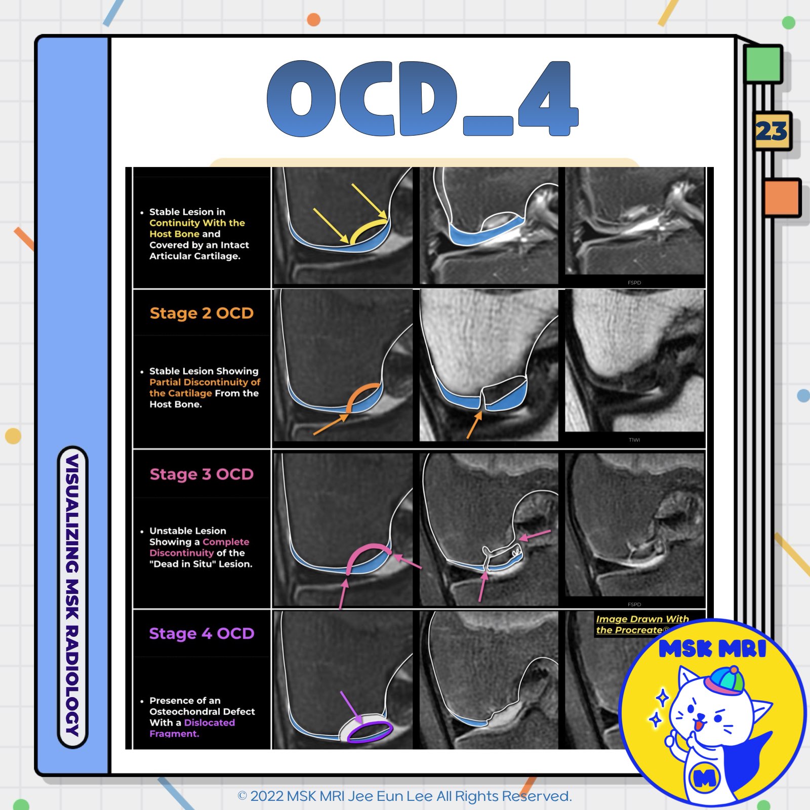
📌ICRS Staging System of Osteochondritis Dissecans
- The International Cartilage Repair Society (ICRS) grading system is adapted for MRI to clinically and arthroscopically evaluate osteochondritis dissecans (OCD).
- As OCD progresses, fragmentation and detachment of the subchondral bone are observed.
- This process of osteochondral fragmentation starts in the deep portion under the articular surface and eventually involves the superficial articular cartilage, suggesting an "inside-out" mechanism.
✅ Stage 1 OCD
- Description: Stable lesion in continuity with the host bone and covered by intact articular cartilage.
- Imaging: The cartilage appears continuous with the bone, and no significant fragmentation is visible.
✅ Stage 2 OCD
- Description: Stable lesion showing partial discontinuity of the cartilage from the host bone.
- Imaging: Partial separation of the cartilage from the underlying bone is observed, indicating some degree of instability.
✅ Stage 3 OCD
- Description: Unstable lesion showing complete discontinuity of the "dead in situ" lesion. However, the fragment is not dislocated.
- Imaging: A clear gap between the lesion and the host bone, but the fragment remains in place.
✅ Stage 4 OCD
- Description: Presence of an osteochondral defect with a dislocated fragment.
- Imaging: The lesion is completely separated from the host bone, with the fragment displaced.
References
- J Clin Orthop Trauma. 2021 Sep 25;22:101610
- Japanese Journal of Radiology (2022) 40:443–457
"Visualizing MSK Radiology: A Practical Guide to Radiology Mastery"
© 2022 MSK MRI Jee Eun Lee All Rights Reserved.
No unauthorized reproduction, redistribution, or use for AI training.
#ICRS #OCD #OsteochondritisDissecans #Radiology #MRI #Arthroscopy #CartilageRepair #BoneFragmentation #MedicalImaging #Orthopedics


'✅ Knee MRI Mastery > Chap 5AB. Chondral and osteochondral' 카테고리의 다른 글
| (Fig 5-B.25) Signs of Osteochondral Lesion Instability in Juveniles (0) | 2024.07.13 |
|---|---|
| (Fig 5-B.24) Signs of Osteochondral Lesion Instability in Adults (0) | 2024.07.13 |
| (Fig 5-B.22) MRI Findings of Osteochondritis Dissecans (0) | 2024.07.13 |
| (Fig 5-B.21) Radiographic Findings of Osteochondritis Dissecans (1) | 2024.07.13 |
| (Fig 5-B.20) Pathogenesis of Osteochondritis Dissecans (0) | 2024.07.13 |