👉 Click the link below and request access—I’ll approve it for you shortly!
https://www.notion.so/MSKMRI-KNEE-b6cbb1e1bc4741b681ecf6a40159a531?pvs=4
==============================================
✨ Join the channel to enjoy the benefits! 🚀
https://www.youtube.com/channel/UC4bw7o0l2rhxn1GJZGDmT9w/join
==============================================
👉 "Click the link to purchase on Amazon 🎉📚"
[Visualizing MSK Radiology: A Practical Guide to Radiology Mastery]
https://www.amazon.com/dp/B0DJGMHMFS
==============================================
MSK MRI Jee Eun Lee
📚 Visualizing MSK Radiology: A Practical Guide to Radiology Mastery Now! 🌟 Available on Amazon, eBay, and Rain Collectibles! 💻 Ebook coming soon – stay tuned! ⏳ 🔗 https://www.amazon.com/dp/B0DJGMHMFS 🔗 https://www.ebay.com/itm/3875004193
www.youtube.com
Visualizing MSK Radiology: A Practical Guide to Radiology Mastery
www.amazon.com
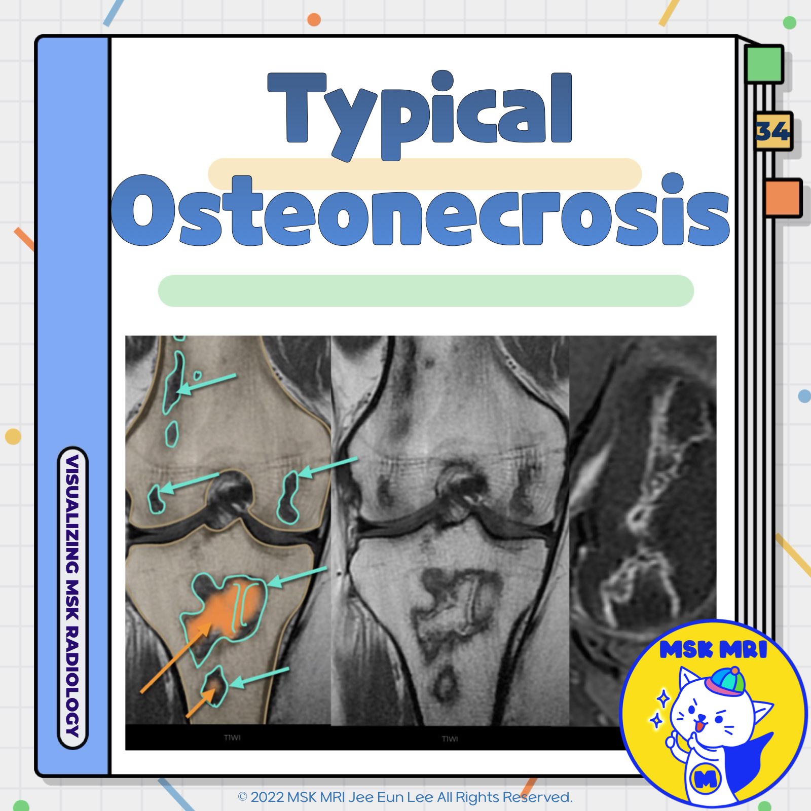
📌 Osteonecrosis
- Osteonecrosis is an umbrella term that includes bone infarcts (metaphyseal) and avascular necrosis (epiphyseal).
- The term "osteonecrosis" is preferred for describing regions of devitalized bone because it is more inclusive than "avascular necrosis" and because all necrosis is by definition avascular.
✅ Double-Line Sign
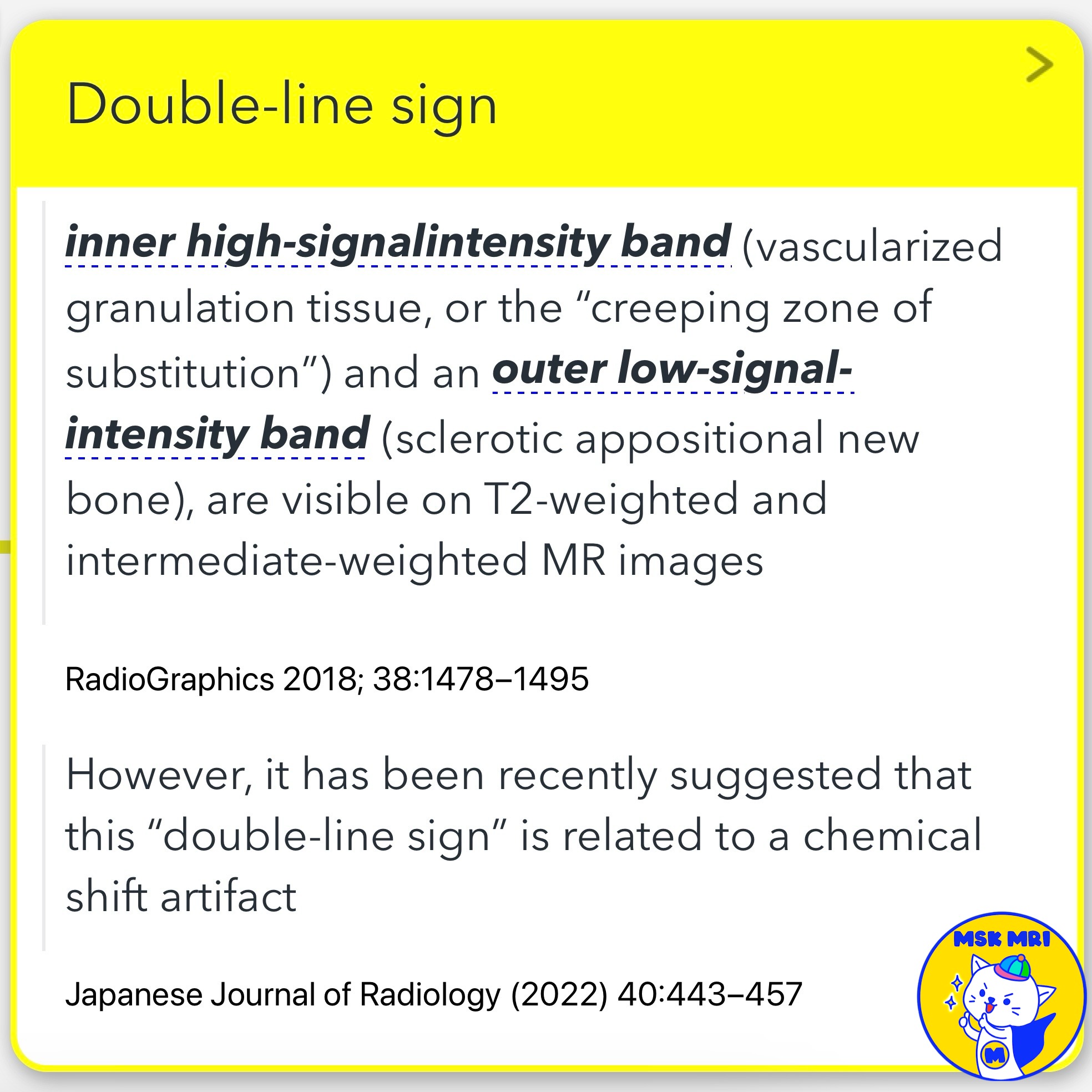
- The double-line sign is seen on T2-weighted and intermediate-weighted MR images.
- It consists of an inner high-signal intensity band (vascularized granulation tissue, or the “creeping zone of substitution”) and an outer low-signal intensity band (sclerotic appositional new bone).
- It has been identified in 65–85% of patients and is considered pathognomonic of osteonecrosis.
- However, recent suggestions indicate that this double-line sign may be related to a chemical shift artifact.
- Histologic Characteristics: Chronic or healing phase osteonecrosis occurs in three zones:
- This rim at the epiphysis represents reparative tissue formed around a crescentic, wedge-shaped, or ringlike region of osteonecrosis.
✅ Mummified fat or yellow marrow
- Regions of osteonecrosis typically consist of central mummified fat or yellow marrow outlined by a distinct uninterrupted rim of primarily low signal intensity.
✅ Early Uncomplicated Osteonecrosis
- The earliest finding of avascular necrosis (AVN) in the hip and knee is a subchondral wedge-shaped, crescent-shaped, or roundish area bordered by a band-like zone of low T1-weighted signal intensity.
References
- AJR 2019; 213:963–982
- RadioGraphics 2018; 38:1478–1495
- Japanese Journal of Radiology (2022) 40:443–457
- Semin Musculoskelet Radiol. 2023 Feb;27(1):45-53
"Visualizing MSK Radiology: A Practical Guide to Radiology Mastery"
© 2022 MSK MRI Jee Eun Lee All Rights Reserved.
No unauthorized reproduction, redistribution, or use for AI training.
#Osteonecrosis, #BoneInfarcts, #AvascularNecrosis, #DoubleLineSign, #MRI, #HistologicCharacteristics, #ChronicOsteonecrosis, #HealingPhase, #EarlyAVN, #Radiology


