👉 Click the link below and request access—I’ll approve it for you shortly!
https://www.notion.so/MSKMRI-KNEE-b6cbb1e1bc4741b681ecf6a40159a531?pvs=4
==============================================
✨ Join the channel to enjoy the benefits! 🚀
https://www.youtube.com/channel/UC4bw7o0l2rhxn1GJZGDmT9w/join
==============================================
👉 "Click the link to purchase on Amazon 🎉📚"
[Visualizing MSK Radiology: A Practical Guide to Radiology Mastery]
https://www.amazon.com/dp/B0DJGMHMFS
==============================================
MSK MRI Jee Eun Lee
📚 Visualizing MSK Radiology: A Practical Guide to Radiology Mastery Now! 🌟 Available on Amazon, eBay, and Rain Collectibles! 💻 Ebook coming soon – stay tuned! ⏳ 🔗 https://www.amazon.com/dp/B0DJGMHMFS 🔗 https://www.ebay.com/itm/3875004193
www.youtube.com
Visualizing MSK Radiology: A Practical Guide to Radiology Mastery
www.amazon.com

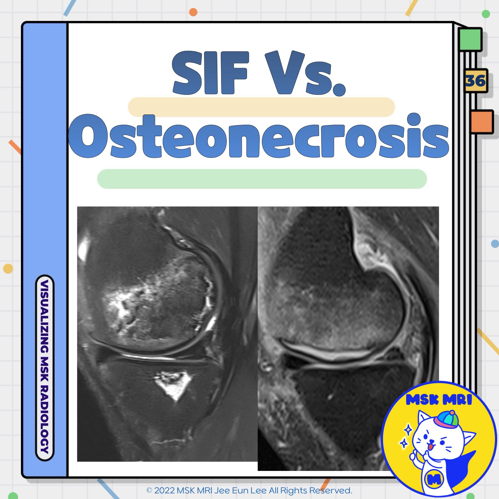
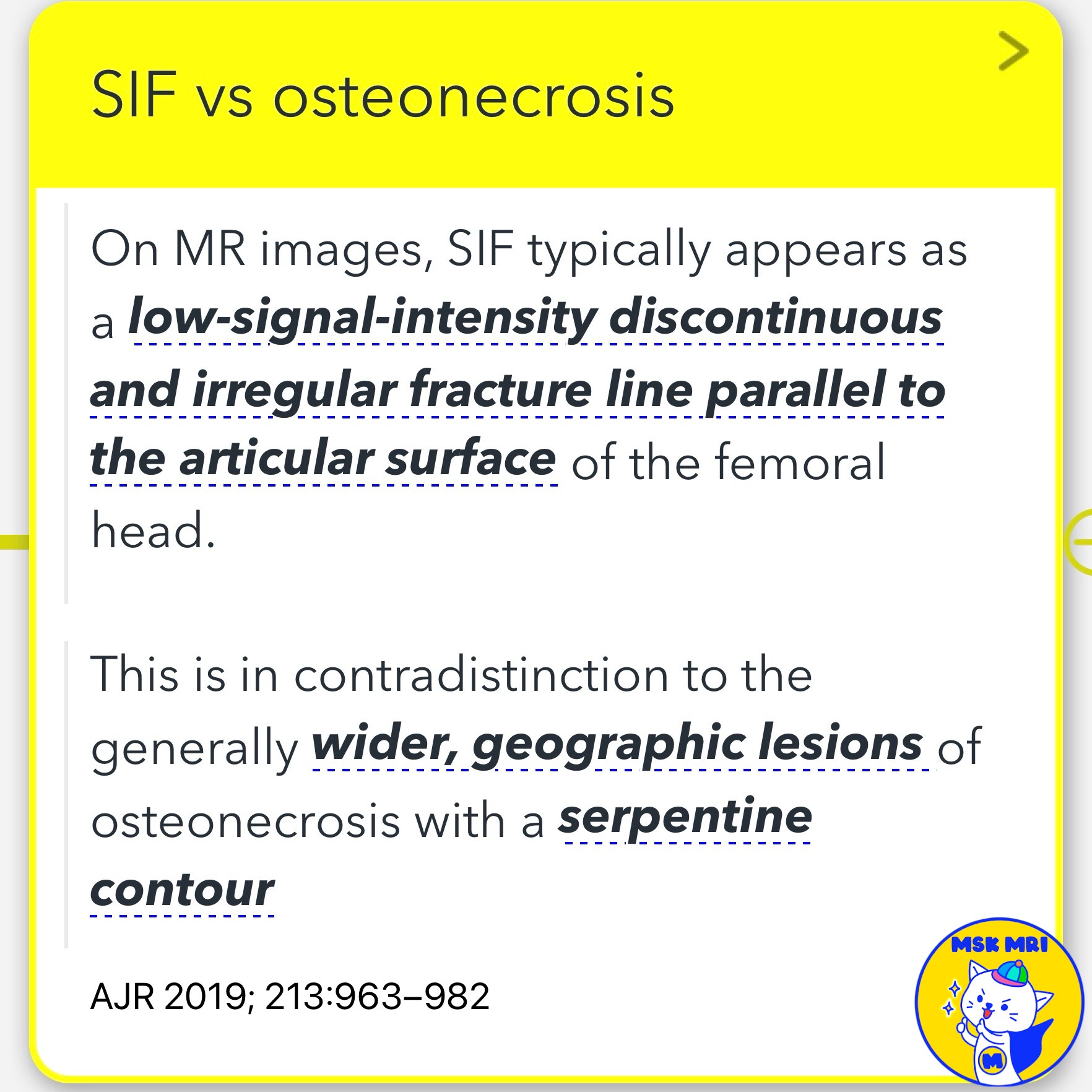
📌 Subchondral Insufficiency Fracture (SIF) vs. Osteonecrosis
✅ SIF on MRI
- Low-signal-intensity discontinuous and irregular fracture line
- Parallel to the articular surface of the femoral head
✅ Osteonecrosis on MRI
- Wider, geographic lesions
- Serpentine contour
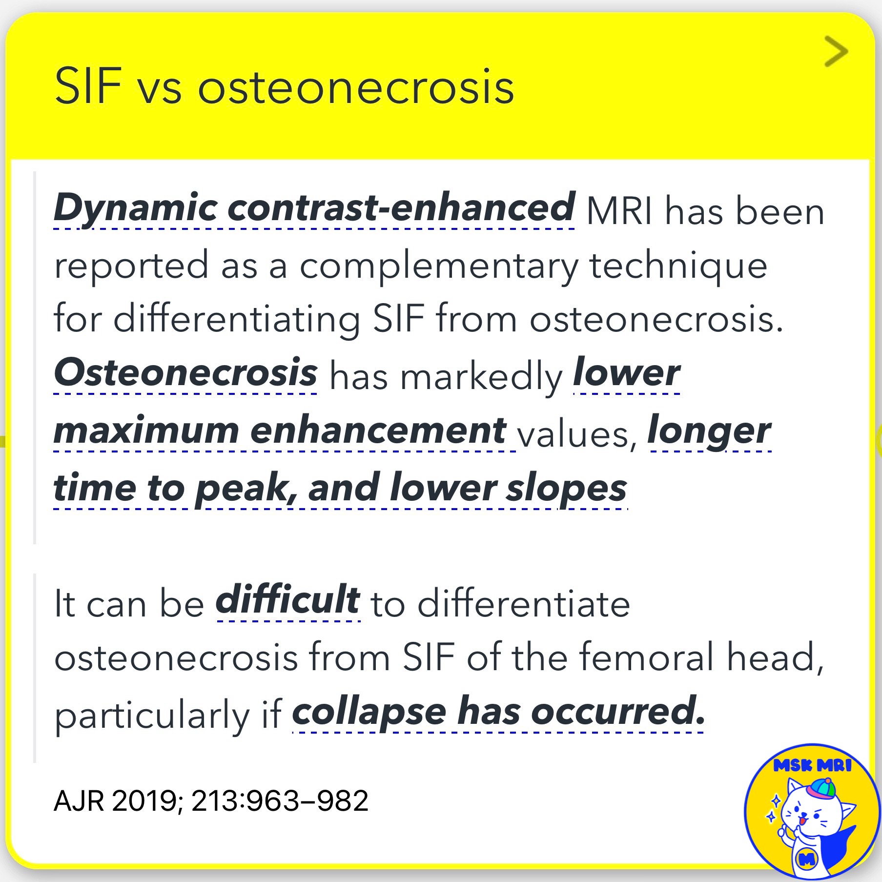
✅ Dynamic Contrast-Enhanced MRI
- Osteonecrosis: Lower maximum enhancement values, longer time to peak, lower slopes
- SIF: Enhancing subchondral portion proximal to the fracture plane (indicating perfused, living bone)
- Osteonecrosis: Non-enhancing regions of dead bone
✅ Importance of Differentiation
- Critical before femoral head collapse
- Osteonecrosis: Considered irreversible
- SIF: May resolve with conservative management
✅ Challenging Cases
- Differentiating SIF without underlying osteonecrosis from SIF with underlying osteonecrosis
- Contrast-enhanced studies may be helpful
References: AJR 2019; 213:963–982
"Visualizing MSK Radiology: A Practical Guide to Radiology Mastery"
© 2022 MSK MRI Jee Eun Lee All Rights Reserved.
No unauthorized reproduction, redistribution, or use for AI training.
Hashtags: #SubchondralInsufficiencyFracture, #Osteonecrosis, #MRI, #FemoralHead, #DynamicContrastEnhancedMRI, #Radiology, #Orthopedics, #BoneImaging, #DifferentialDiagnosis, #ConservativeManagement


'✅ Knee MRI Mastery > Chap 5AB. Chondral and osteochondral' 카테고리의 다른 글
| (Fig 5-B.35) Patterns of Subchondral Bone Plate Fracture in Osteonecrosis (0) | 2024.07.14 |
|---|---|
| (Fig 5-B.34) Typical Osteonecrosis (0) | 2024.07.14 |
| (Fig 5-B.33) Poor Prognostic Factors in Subchondral Insufficiency Fracture (0) | 2024.07.14 |
| (Fig 5-B.32) Subchondral Fracture with Epiphyseal Collapse: Part 2 (0) | 2024.07.14 |
| (Fig 5-B.31) Subchondral Fracture with Epiphyseal Collapse: Part 1 (0) | 2024.07.14 |