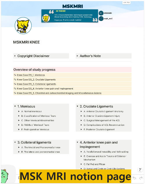
👉 Click the link below and request access—I’ll approve it for you shortly!
https://www.notion.so/MSKMRI-KNEE-b6cbb1e1bc4741b681ecf6a40159a531?pvs=4
==============================================
✨ Join the channel to enjoy the benefits! 🚀
https://www.youtube.com/channel/UC4bw7o0l2rhxn1GJZGDmT9w/join
==============================================
👉 "Click the link to purchase on Amazon 🎉📚"
[Visualizing MSK Radiology: A Practical Guide to Radiology Mastery]
https://www.amazon.com/dp/B0DJGMHMFS
==============================================
MSK MRI Jee Eun Lee
📚 Visualizing MSK Radiology: A Practical Guide to Radiology Mastery Now! 🌟 Available on Amazon, eBay, and Rain Collectibles! 💻 Ebook coming soon – stay tuned! ⏳ 🔗 https://www.amazon.com/dp/B0DJGMHMFS 🔗 https://www.ebay.com/itm/3875004193
www.youtube.com
Visualizing MSK Radiology: A Practical Guide to Radiology Mastery
www.amazon.com
📌 Common Complications Post-Autologous Chondrocyte Implantation (ACI)
- Three primary complications are typically observed on MRI following ACI:
✅ Graft Hypertrophy:
- This occurs when the repair tissue is thicker than the native cartilage, reported in 10 to 63% of cases.
- Although often asymptomatic, severe hypertrophy can cause symptomatic "catching" and may require resection.
- Kreuz et al. found that all patients with high-grade hypertrophy needed revision shaving.
✅ Graft Failure:
- Assessment of graft incorporation is based on tissue continuity between the graft and host cartilage.
- Issues such as fissuring at the interface, fluid between graft and host bone, and subchondral cyst formation indicate incomplete integration or failure of the graft.
- Manifested as a full-thickness defect or absence of the graft.
- Degeneration, fraying, and delamination may also occur, potentially necessitating revision or repair.
✅ Arthrofibrosis:
- Reported in 5 to 10% of cases, this is seen as a dark signal band in Hoffa’s fat pad or focal thickening of the joint capsule, leading to clinical restriction.
- It is more common in patients with extensive surgical histories and can be diagnosed clinically or via MRI.
✅ MOCART 2.0 Knee Score
- The MOCART 2.0 knee score is an updated version of the original MOCART score, reflecting advancements in surgical treatments for cartilage defects and their MRI appearances.
Volume Fill of Cartilage Defect:
- Complete filling or minor hypertrophy (100% to 150%): 20 points
- Major hypertrophy (≥150%) or 75% to 99% filling: 15 points
- 50% to 74% filling: 10 points
- 25% to 49% filling: 5 points
- <25% filling or complete delamination in situ: 0 points
References
- Semin Musculoskelet Radiol 2011;15:69–88
- J Clin Orthop Trauma. 2021 Sep 25;22:101610
"Visualizing MSK Radiology: A Practical Guide to Radiology Mastery"
© 2022 MSK MRI Jee Eun Lee All Rights Reserved.
No unauthorized reproduction, redistribution, or use for AI training.
#AutologousChondrocyteImplantation, #CartilageRepair, #GraftHypertrophy, #GraftFailure, #Arthrofibrosis, #MOCARTScore, #KneeMRI, #OrthopedicSurgery, #CartilageDefects, #SportsMedicine


