👉 Click the link below and request access—I’ll approve it for you shortly!
https://www.notion.so/MSKMRI-KNEE-b6cbb1e1bc4741b681ecf6a40159a531?pvs=4
==============================================
✨ Join the channel to enjoy the benefits! 🚀
https://www.youtube.com/channel/UC4bw7o0l2rhxn1GJZGDmT9w/join
==============================================
👉 "Click the link to purchase on Amazon 🎉📚"
[Visualizing MSK Radiology: A Practical Guide to Radiology Mastery]
https://www.amazon.com/dp/B0DJGMHMFS
==============================================
MSK MRI Jee Eun Lee
📚 Visualizing MSK Radiology: A Practical Guide to Radiology Mastery Now! 🌟 Available on Amazon, eBay, and Rain Collectibles! 💻 Ebook coming soon – stay tuned! ⏳ 🔗 https://www.amazon.com/dp/B0DJGMHMFS 🔗 https://www.ebay.com/itm/3875004193
www.youtube.com
Visualizing MSK Radiology: A Practical Guide to Radiology Mastery
www.amazon.com

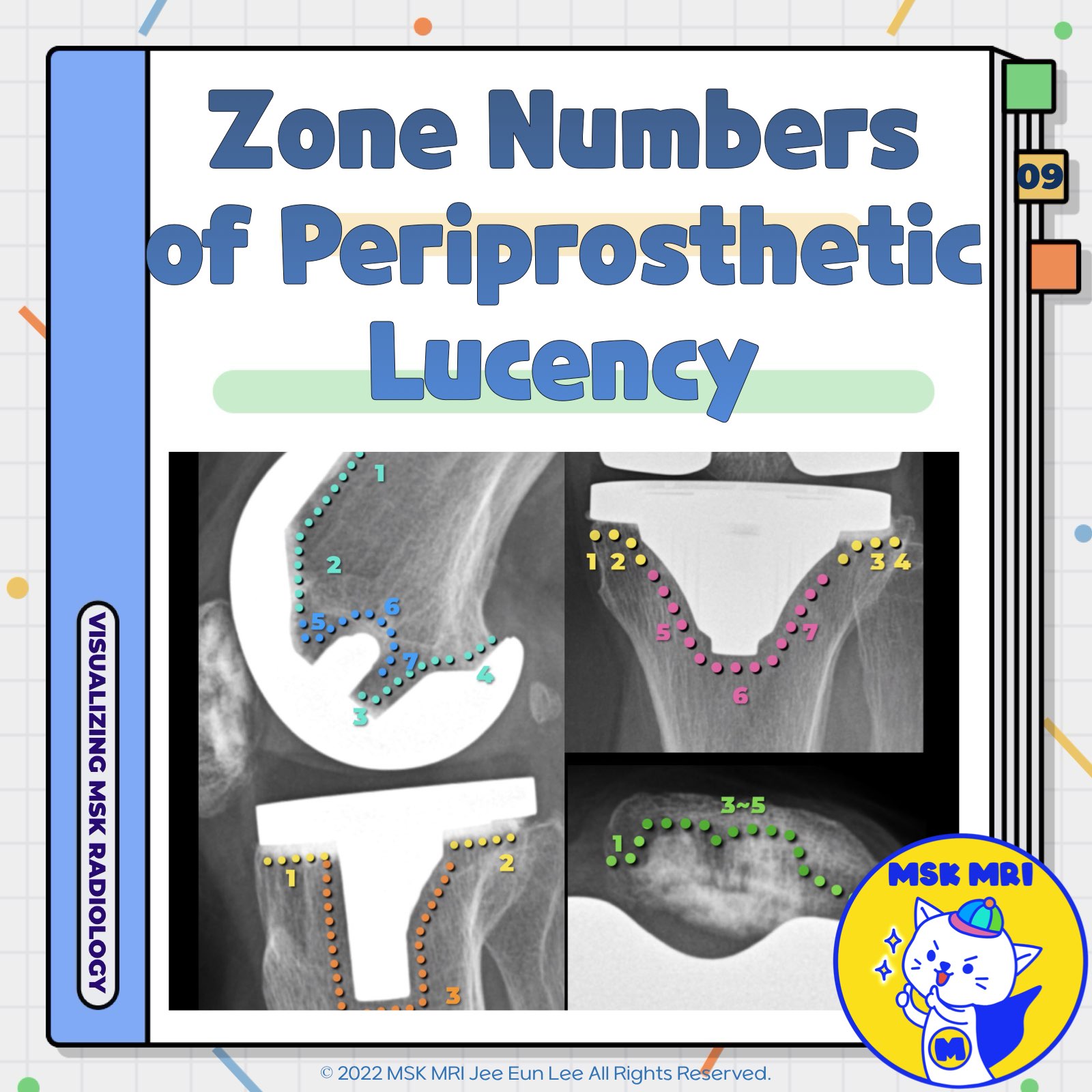
📌 Specific Zone Numbers of Periprosthetic Lucency after TKA
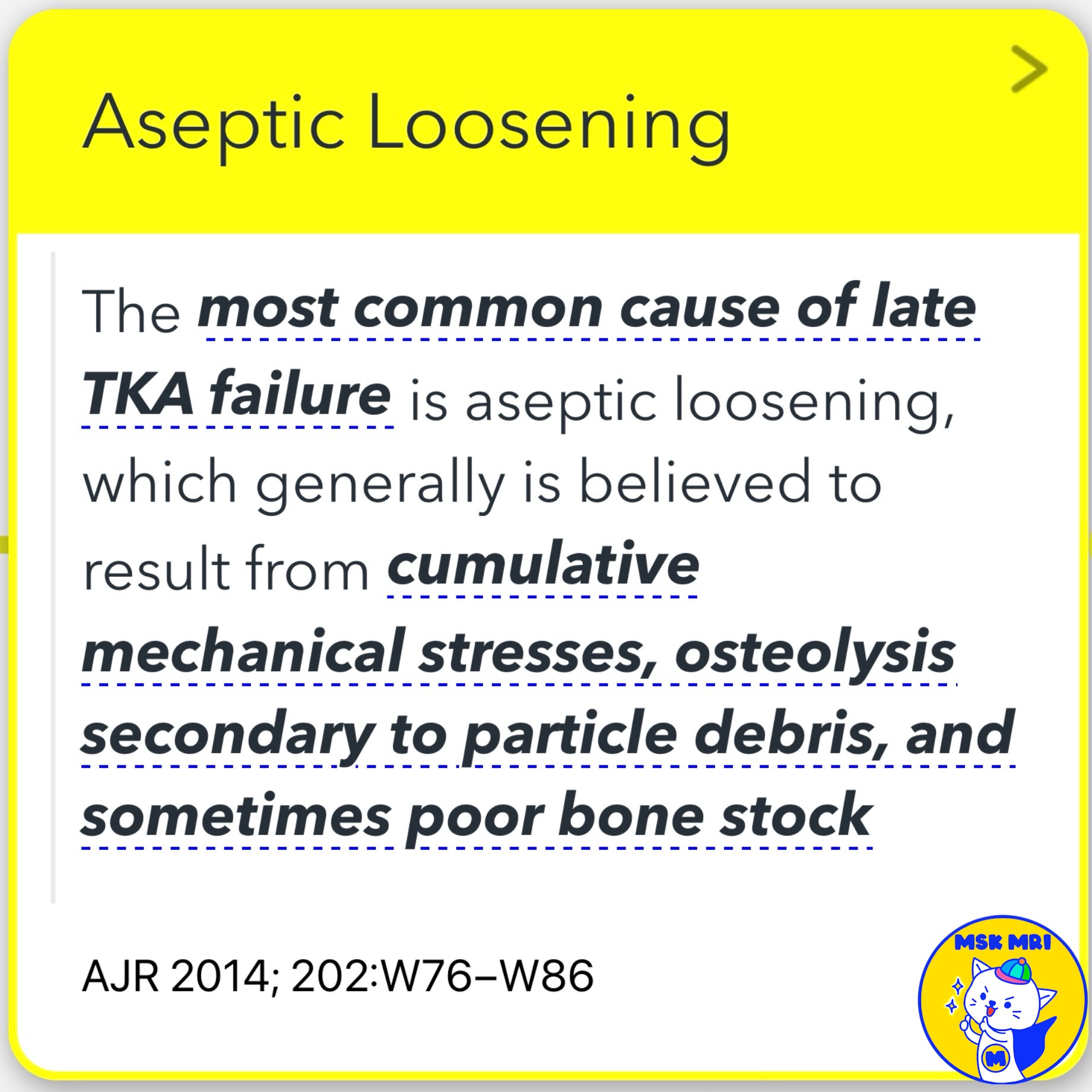
✅ Aseptic Loosening
- The most common cause of late TKA failure is aseptic loosening, which generally is believed to result from cumulative mechanical stresses, osteolysis secondary to particle debris, and sometimes poor bone stock.
- Radiographic criteria for loosening include wide or progressively enlarging radiolucency at the cement-bone, metal-cement, or metal-bone interface; component migration or subsidence; and cement fractures.
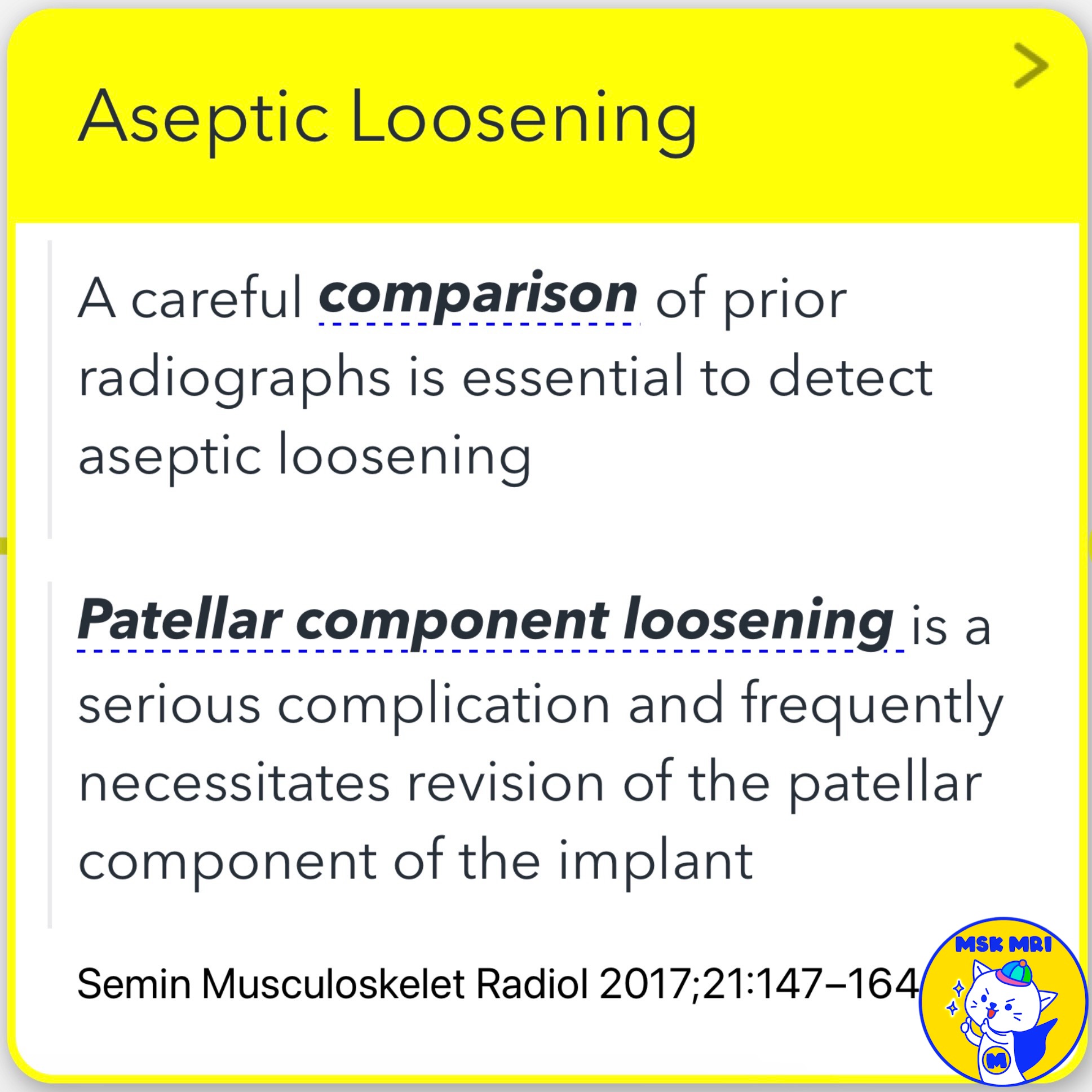
- A careful comparison of prior radiographs is essential to detect aseptic loosening.
- Patellar component loosening is a serious complication that frequently necessitates revision of the implant's patellar component.
- Radiographic findings of aseptic loosening include periprosthetic lucency between the bone–cement or metal–bone interface usually greater than 2 mm.
- Increases in the width of the periprosthetic radiolucency, focal radiolucency greater than 2 mm, and component migration indicate loosening.
✅ Periprosthetic Lucency Evaluation
- The “Knee Society” has formulated a standardized grading system by ascribing specific zone numbers to the periprosthetic interfaces, thereby standardizing the grading and description of these lucent zones.
References
- AJR 2014; 202:W76–W86
- Semin Musculoskelet Radiol 2017; 21:147–164
- Clin Orthop Relat Res 1989; 248:9–12
"Visualizing MSK Radiology: A Practical Guide to Radiology Mastery"
© 2022 MSK MRI Jee Eun Lee All Rights Reserved.
No unauthorized reproduction, redistribution, or use for AI training.
#AsepticLoosening, #TKA, #RadiographicCriteria, #KneeSociety, #PeriprostheticLucency, #Radiolucency, #ComponentMigration, #BoneCementInterface, #PatellarComponent, #OrthopedicRadiology


'✅ Knee MRI Mastery > Chap 5CD. Cartilage Repair and TKA' 카테고리의 다른 글
| (Fig 5-D.11) Stress Shielding (0) | 2024.07.19 |
|---|---|
| (Fig 5-D.10) Aseptic Loosening (0) | 2024.07.19 |
| (Fig 5-D.08) Dislocation of Polyethylene Insert (0) | 2024.07.19 |
| (Fig 5-D.07) Polyethylene Wear-Induced Synovitis and Osteolysis (0) | 2024.07.19 |
| (Fig 5-D.06) Polyethylene Wear–Induced Synovitis (0) | 2024.07.19 |