📌 Calcaneal Intraosseous Lipoma
✅ Diagnostic Features
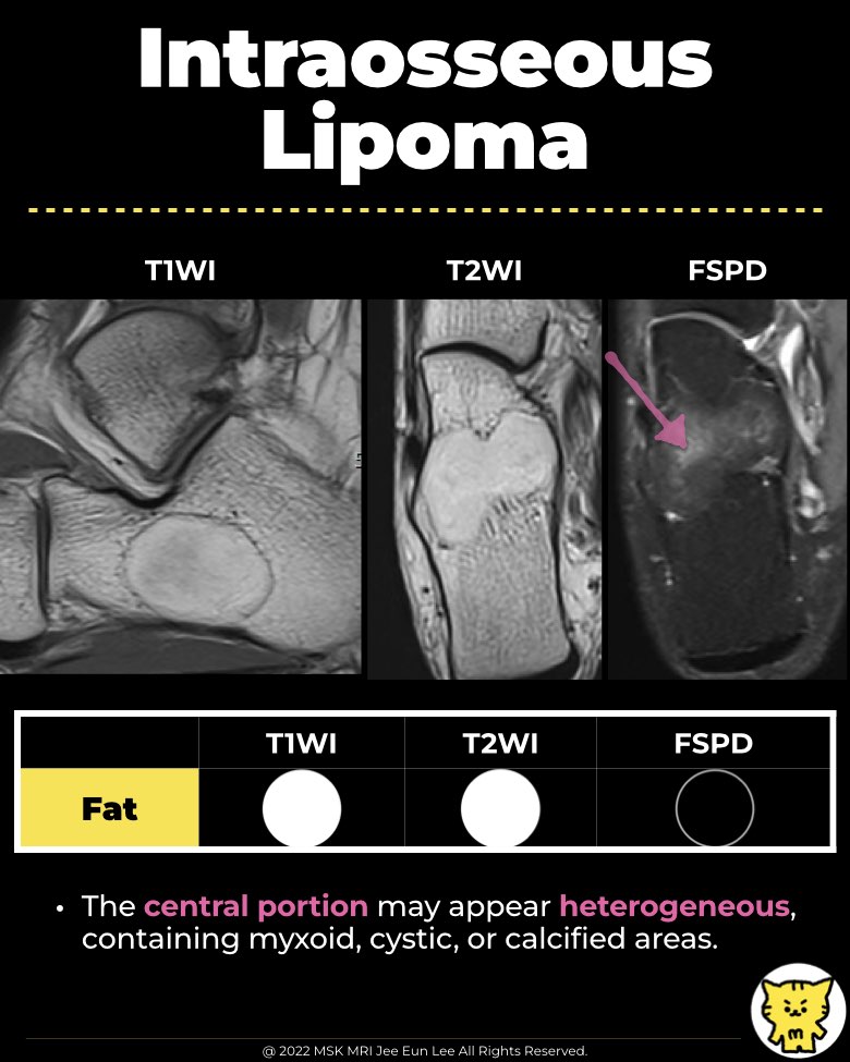
- Obvious fat signal intensity on T1-weighted MRI images
- High sensitivity and specificity for diagnosis
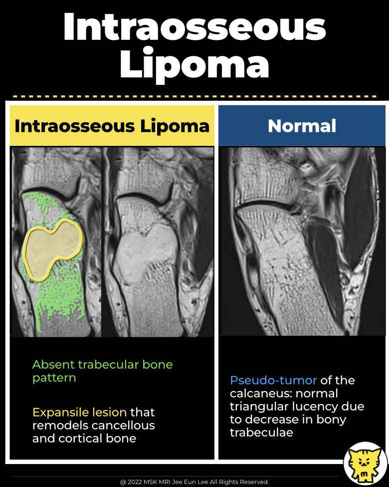
- Expansile lesion that remodels cancellous and cortical bone (distinguishes from bone infarction)
✅ Differential Diagnosis
- Bone infarct
- Unicameral bone cyst
- Aneurysmal bone cyst
- Chondromyxoid fibroma
- Osteoblastoma
- Giant cell tumor
✅ Unicameral bone cyst vs intraosseous lipoma
- Bone Cyst:
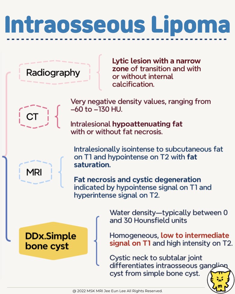
- Appears as a water-density lesion (0–30 Hounsfield units).
- Low signal intensity on T1-weighted images.
- High signal intensity on T2-weighted images.
- Does not enhance after contrast administration.
- Intraosseous Lipoma:
- High signal intensity on T1-weighted images.
- Relatively high signal intensity on T2-weighted images.
- Very low intensity on fat-suppressed sequences.
- May show a slightly higher central signal due to granulation tissue or necrosis.
#IntraosseousLipomas, #BoneTumors, #BenignLesions, #LipogenicTumors, #Calcaneus, #BoneCyst, #IntraosseousLipoma, #Radiology, #MusculoskeletalImaging, #Radiolucency, #Pseudolesion
"Visualizing MSK Radiology: A Practical Guide to Radiology Mastery"
© 2022 MSK MRI Jee Eun Lee All Rights Reserved.
No unauthorized reproduction, redistribution, or use for AI training.