✨ Join the channel to enjoy the benefits! 🚀
https://www.youtube.com/channel/UC4bw7o0l2rhxn1GJZGDmT9w/join
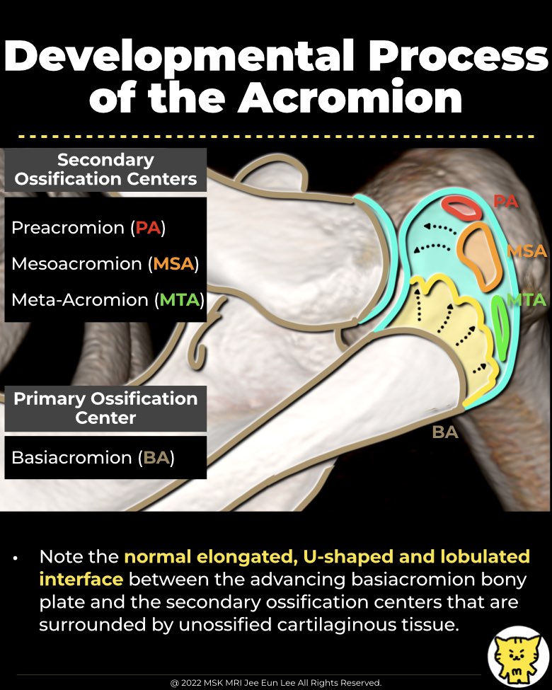
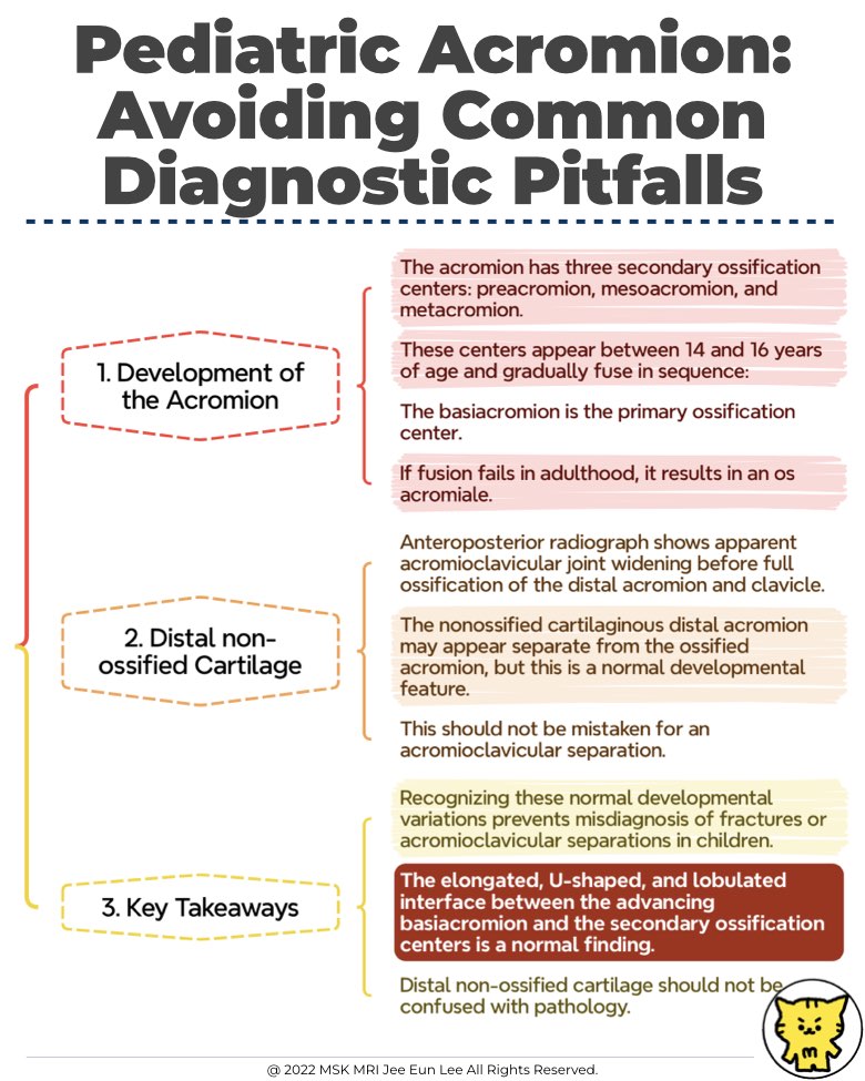
1. Development of the Acromion
- The acromion has three secondary ossification centers: preacromion, mesoacromion, and metacromion.
- These centers appear between 14 and 16 years of age and gradually fuse in sequence:
- The basiacromion is the primary ossification center.
- If fusion fails in adulthood, it results in an os acromiale.
2. Normal Radiographic Findings in Children
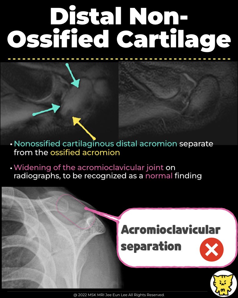
- In pediatric patients, the distal acromion and lateral clavicular epiphysis are partially unossified, which can mimic acromioclavicular joint widening or fractures.
- The nonossified cartilaginous distal acromion may appear separate from the ossified acromion, but this is a normal developmental feature.
3. Case Example: 8-Year-Old Boy
- Anteroposterior radiograph shows apparent acromioclavicular joint widening before full ossification of the distal acromion and clavicle.
- This should not be mistaken for an acromioclavicular separation.
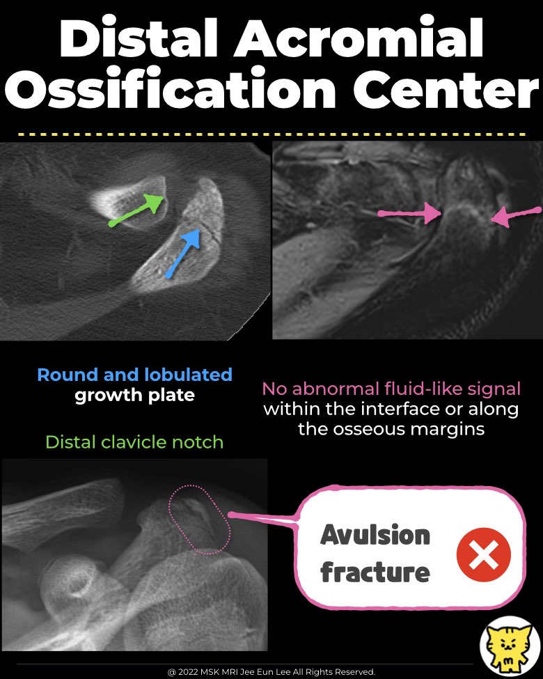
4. Normal Pediatric Acromion: A Closer Look
- Radiographs of a 13-year-old boy and a 12-year-old girl show normally developing acromial ossification centers.
- The elongated, U-shaped, and lobulated interface between the advancing basiacromion and the secondary ossification centers is a normal finding.
- The distal clavicular notch, a temporary developmental feature, disappears once ossification is complete.
5. Key Takeaways
- Recognizing these normal developmental variations prevents misdiagnosis of fractures or acromioclavicular separations in children.
- Distal non-ossified cartilage should not be confused with pathology.
#PediatricRadiology, #ShoulderImaging, #AcromionDevelopment, #OssificationCenters, #OsAcromiale, #XrayFindings, #MSKRadiology, #AcromioclavicularJoint, #FractureMimics, #RadiologyEducation
"Visualizing MSK Radiology: A Practical Guide to Radiology Mastery"
© 2022 MSK MRI Jee Eun Lee All Rights Reserved.
No unauthorized reproduction, redistribution, or use for AI training.