✨ Join the channel to enjoy the benefits! 🚀https://www.youtube.com/channel/UC4bw7o0l2rhxn1GJZGDmT9w/join
📌Os Acromiale: Summary and Classification
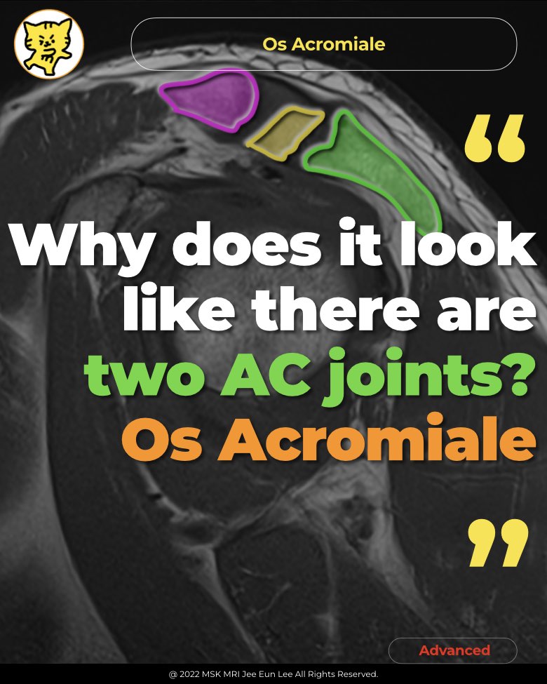
✅Definition
Os acromiale results from the failure of fusion of at least one of the secondary ossification centers of the acromion.
✅ Classification of Os Acromiale
Os acromiale types are classified based on the unfused segment located immediately anterior to the site of nonunion.
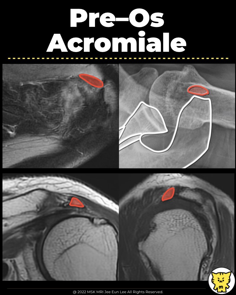
- Pre–os acromiale: Nonunion between the preacromion and mesoacromion.
- Meso–os acromiale: Nonunion between the mesoacromion and meta-acromion.
- Meta–os acromiale: Nonunion between the meta-acromion and basiacromion.
➡️ Most Common Types
- The most frequent os acromiale is the mesoacromion, which has a large, triangular shape and forms an interface near the acromioclavicular joint (ACJ).
- The preacromion, a less common type, is located at the distal tip of the acromion.
- Axillary shoulder radiographs and axial MR images are the best imaging modalities to detect an os acromiale.
✅ Clinical Significance of Os Acromiale
- Os acromiale is often asymptomatic and may be an incidental finding on imaging.
- In some cases, os acromiale can contribute to impingement syndrome and cause pain.
- Pain may result from mechanical instability and pseudarthrosis formation at the site of nonunion.
- Both instability and pseudarthrosis can worsen after acromioplasty, potentially leading to increased symptoms.
✅ References
- Pediatr Radiol. 2021 Mar; 51(3):338-352.
- J Shoulder Elbow Surg. 2020 Feb; 29(2):402-410.
- Radiographics. 2015 Jul-Aug; 35(4):1108-22.
- Pediatr Radiol. 2019 Nov; 49(12):1617-1628.
- J Belg Soc Radiol. 2017 Dec 16; 101(Suppl 2):3.
"Visualizing MSK Radiology: A Practical Guide to Radiology Mastery"
© 2022 MSK MRI Jee Eun Lee All Rights Reserved.
No unauthorized reproduction, redistribution, or use for AI training.