https://youtu.be/P8_SWtmvI9s?si=S0N7FLPsFK6LP48L
✨ Join the channel to enjoy the benefits! 🚀 https://www.youtube.com/channel/UC4bw7o0l2rhxn1GJZGDmT9w/join
👉 Click the link below and request access—I’ll approve it for you shortly! https://www.notion.so/MSKMRI-KNEE-b6cbb1e1bc4741b681ecf6a40159a531?pvs=4
📚 Visualizing MSK Radiology:
A Practical Guide to Radiology Mastery Now!
🌟 Available on Amazon and eBay!
🔗 https://www.amazon.com/dp/B0DJGMHMFS
🔗 https://www.ebay.com/itm/387500419368
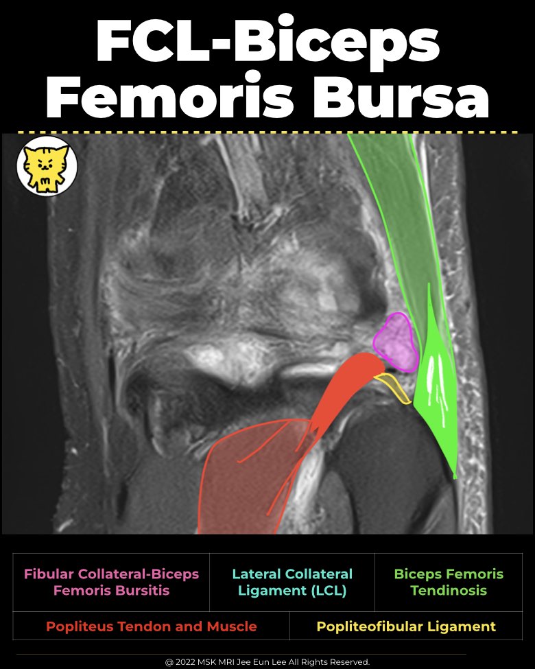
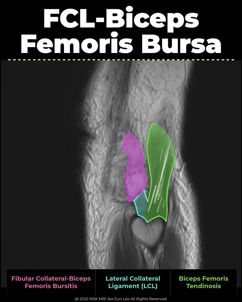
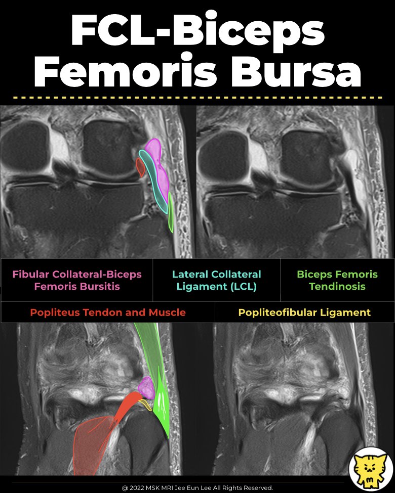
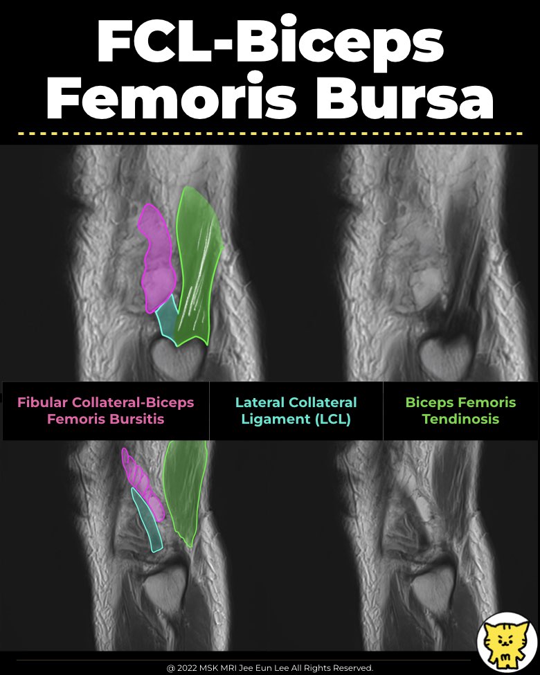
✅ Anatomy and Location
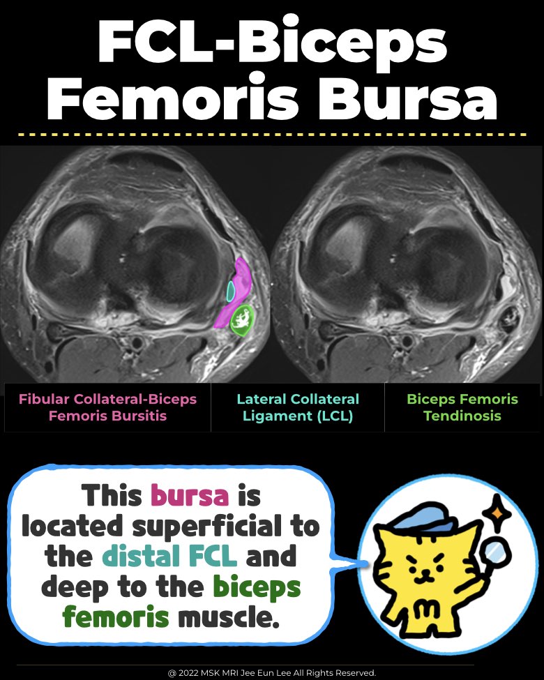
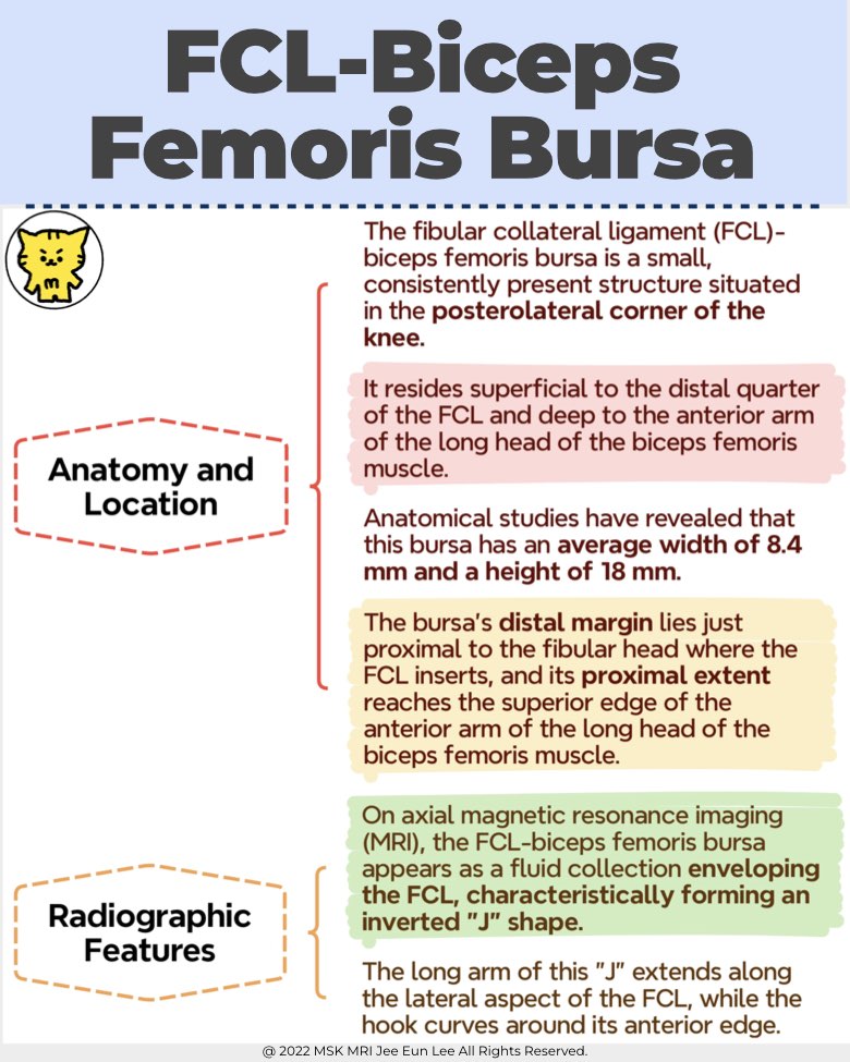
- The fibular collateral ligament (FCL)-biceps femoris bursa is a small, consistently present structure situated in the posterolateral corner of the knee.
- It resides superficial to the distal quarter of the FCL and deep to the anterior arm of the long head of the biceps femoris muscle.
- Anatomical studies have revealed that this bursa has an average width of 8.4 mm and a height of 18 mm.
- The bursa's distal margin lies just proximal to the fibular head where the FCL inserts, and its proximal extent reaches the superior edge of the anterior arm of the long head of the biceps femoris muscle.
✅ Radiographic Features
- On axial magnetic resonance imaging (MRI), the FCL-biceps femoris bursa appears as a fluid collection enveloping the FCL, characteristically forming an inverted "J" shape.
- The long arm of this "J" extends along the lateral aspect of the FCL, while the hook curves around its anterior edge.
- This configuration is crucial for radiologists to recognize, as it aids in distinguishing the bursa from pathological cyst-like structures around the knee.
✅ Clinical Significance
- Important for diagnosing lateral knee pain
- Helps distinguish bursitis from other knee pathologies
- Recognizing prevalence, shape, and size aids in accurate assessment
"Visualizing MSK Radiology: A Practical Guide to Radiology Mastery"
© 2022 MSK MRI Jee Eun Lee All Rights Reserved.
No unauthorized reproduction, redistribution, or use for AI training.
#FCLBursitis, #LateralCollateralLigamentBursitis, #BicepsFemorisBursitis, #KneeBursitis, #KneeAnatomyMRI,
#kneepain, #bicepsfemoristendinosis, #bursitis, #tendonthickening, #tendoninjuries,


'✅ Knee MRI Mastery > Chap 3.Collateral Ligaments' 카테고리의 다른 글
| Lateral Knee Pain? Don’t Ignore Iliotibial Band Syndrome! (0) | 2025.02.01 |
|---|---|
| 📌 Segond Fracture Explained: The Hidden Clue in ACL Tears! (0) | 2025.01.26 |
| Segond Fracture: Key Indicator of ACL Injury You Shouldn’t Miss (0) | 2025.01.15 |
| (Fig 3-B.40, 41) Anterolateral Ligament Reconstruction (0) | 2024.05.25 |
| (Fig 3-B.38, 39, 42) Posterolateral Corner Reconstruction (0) | 2024.05.24 |