https://youtube.com/shorts/6mX5LQ5eJgI
- YouTube
www.youtube.com
 Coalition by MSKMRI JEE EUN LEE.pdf
6.67MB
Coalition by MSKMRI JEE EUN LEE.pdf
6.67MB
The subtalar joint (talocalcaneal joint) is vital for hindfoot stability and mobility.
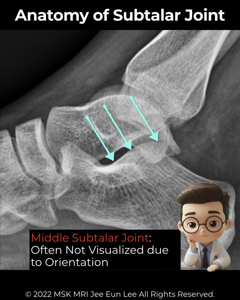
- Posterior facet: largest, best seen on lateral radiographs and CT
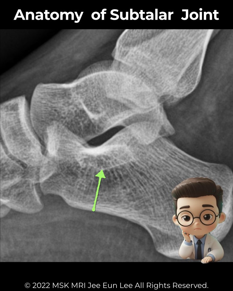
- Middle facet: on the sustentaculum tali, may communicate with the talonavicular joint
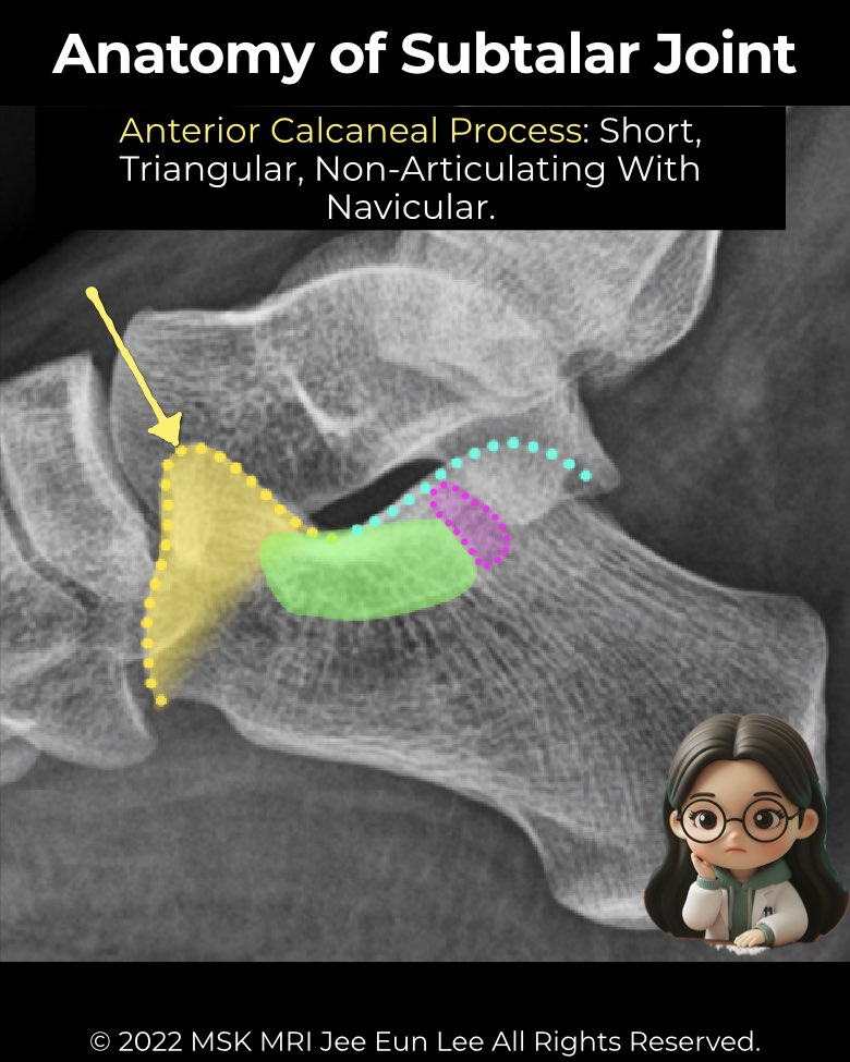
- Anterior facet: smallest, may merge with the middle facet
Imaging pearls:
- Sustentaculum tali appears brick-shaped on lateral X-ray
- Anterior calcaneal process should not articulate with the navicular—if it does, think coalition
- Harris view: posterior and middle facets normally parallel (<20°)
- CT/MRI: gold standard for coalitions and subtle variants
#Radiology, #MSKMRI, #SubtalarJoint, #FootMRI, #TalocalcanealJoint, #OrthopedicImaging, #RadiologyEducation, #MRIteaching, #RadiologistLife, #MedicalEducation
Visualizing MSK Radiology: A Practical Guide to Radiology Mastery
© 2022 MSK MRI Jee Eun Lee All Rights Reserved.
No unauthorized reproduction, redistribution, or use for AI training.