https://youtube.com/shorts/uTfYbMWfHQA
 Coalition by MSKMRI JEE EUN LEE.pdf
6.67MB
Coalition by MSKMRI JEE EUN LEE.pdf
6.67MB
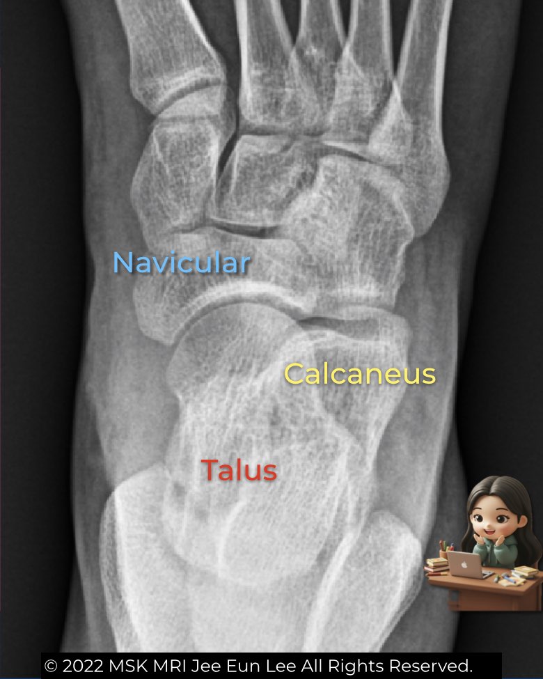
The AP and oblique foot views are essential for assessing alignment and detecting early coalitions.
- AP view: check the smooth alignment between the lateral margin of the talar head and the navicular. Any step-off suggests abnormality. The anterior process of the calcaneus is usually not seen. This view also allows angle measurements like the talar–first metatarsal angle to evaluate flatfoot.
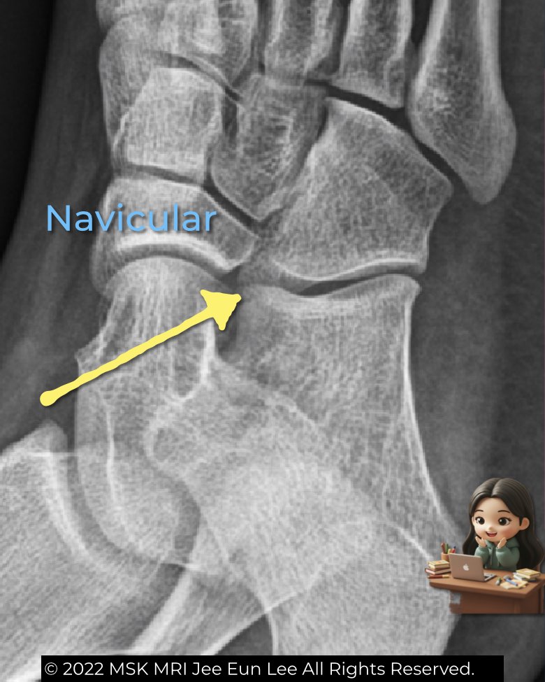
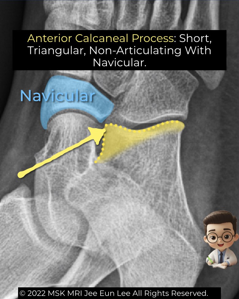
- Oblique view: best for the calcaneonavicular space. Normally, the anterior process of the calcaneus is short, triangular, and separated from the navicular. If it elongates and approaches the navicular, think coalition (Anteater sign).
These simple views provide quick radiographic clues before moving to CT or MRI for confirmation.
#Radiology, #MSKMRI, #FootXray, #CalcaneonavicularCoalition, #AnteaterSign, #OrthopedicImaging, #RadiologyEducation, #MSKImaging, #RadiologistLife, #MRIteaching
Visualizing MSK Radiology: A Practical Guide to Radiology Mastery
© 2022 MSK MRI Jee Eun Lee All Rights Reserved.
No unauthorized reproduction, redistribution, or use for AI training.