https://youtube.com/shorts/k-R-P-7OiJY
 Coalition by MSKMRI JEE EUN LEE.pdf
6.67MB
Coalition by MSKMRI JEE EUN LEE.pdf
6.67MB
The subtalar joint is a complex articulation between the talus and calcaneus, best assessed with MRI for both normal anatomy and coalition-related pathology.
Talocalcaneal articulations
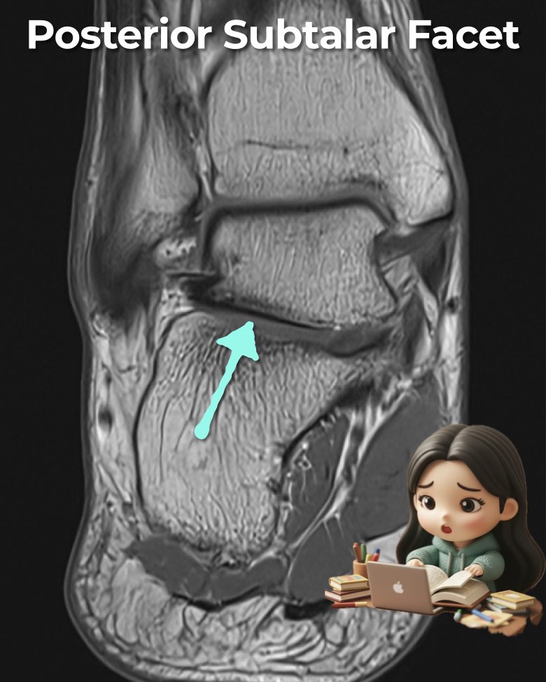
- Posterior subtalar joint: concave talar facet with convex calcaneal facet, largest and best seen in coronal/sagittal planes.
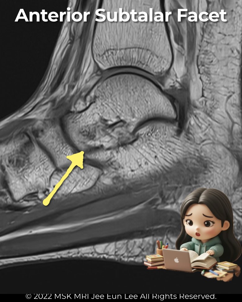
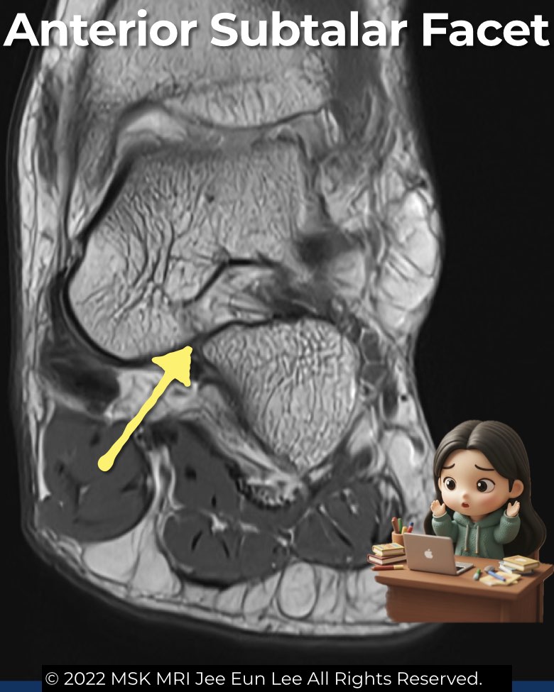
- Talocalcaneonavicular joint: talar head with navicular, plus anterior and middle facets.
Articular facets on MRI
- Posterior facet: largest, clearly seen on coronal, sagittal, and axial sequences.
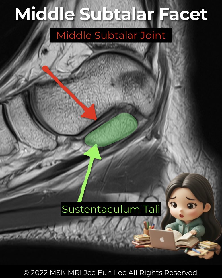
- Middle facet: sustentaculum tali with talar head; normally horizontal/upward, but downward medial slope = “drunken waiter sign” → middle facet coalition. Sustentaculum appears brick-shaped.
- Anterior facet: smallest, variably present, anterior-lateral to the middle facet.
Tarsal canal & sinus tarsi
- Canal separates posterior from anterior/middle joints.
- MRI shows sinus tarsi as a fat-filled cone; edema here is strongly associated with subtalar pathology.
Ligaments on MRI
- Interosseous talocalcaneal ligament (ITCL): stout band in the tarsal canal, best on sagittal/coronal PD.
- Anterior capsular ligament: capsular thickening, just posterior-lateral to ITCL.
- Medial talocalcaneal ligament: variant, from talar process to sustentaculum; smooth attachments distinguish it from fibrous coalition.
Normal anatomic variants
- Conjoined anterior and middle facets are most common (53–62%).
- Anterior facet may be absent or continuous with other facets.
- Accessory facets (posterior sustentaculum ↔ talar process): smooth cortical margins = normal variant, not coalition.
#Radiology, #MSKMRI, #SubtalarJoint, #FootMRI, #CoalitionImaging, #OrthopedicImaging, #RadiologyEducation, #MSKImaging, #RadiologistLife, #MRIteaching
Visualizing MSK Radiology: A Practical Guide to Radiology Mastery
© 2022 MSK MRI Jee Eun Lee All Rights Reserved.
No unauthorized reproduction, redistribution, or use for AI training.