https://youtube.com/shorts/f7KNKr3Yfpg
 Coalition by MSKMRI JEE EUN LEE.pdf
6.67MB
Coalition by MSKMRI JEE EUN LEE.pdf
6.67MB
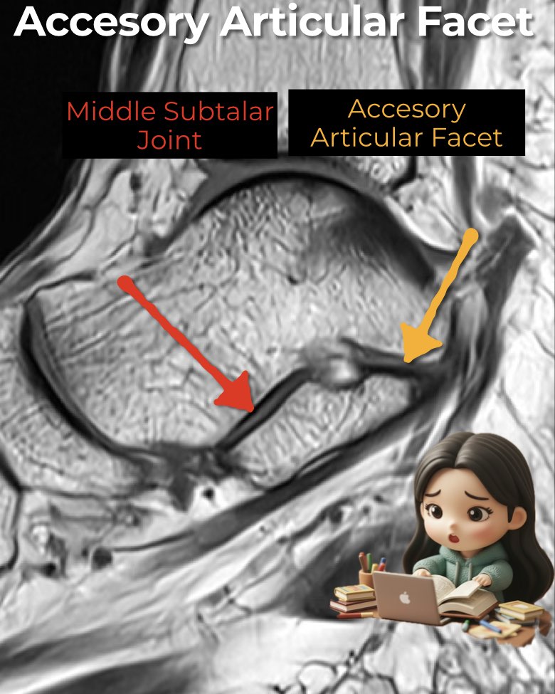
Accessory articular facets are normal anatomic variants of the talocalcaneal joint but can mimic or associate with pathology.
Accessory Anterolateral Talar Facet (AALTF):
- Extension of posterior talar facet cartilage, seen anterior to the lateral talar process.
- Prevalence: ~3–10%.
- MRI findings: strongly associated with focal abutting bone marrow edema (FABME), sinus tarsi edema, and subtalar irritation (may cause peroneal spastic flatfoot).
- Coalition link: up to 29% of patients with AALTF also have a tarsal coalition, most often talocalcaneal (especially extra-articular or middle facet). Always search for both.
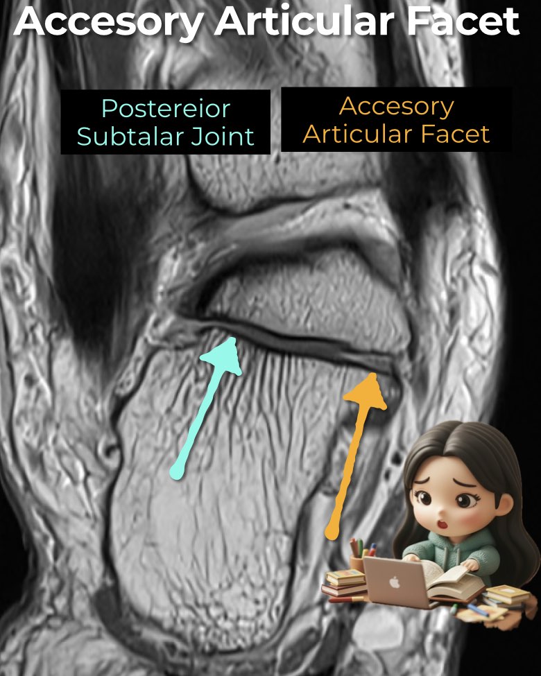
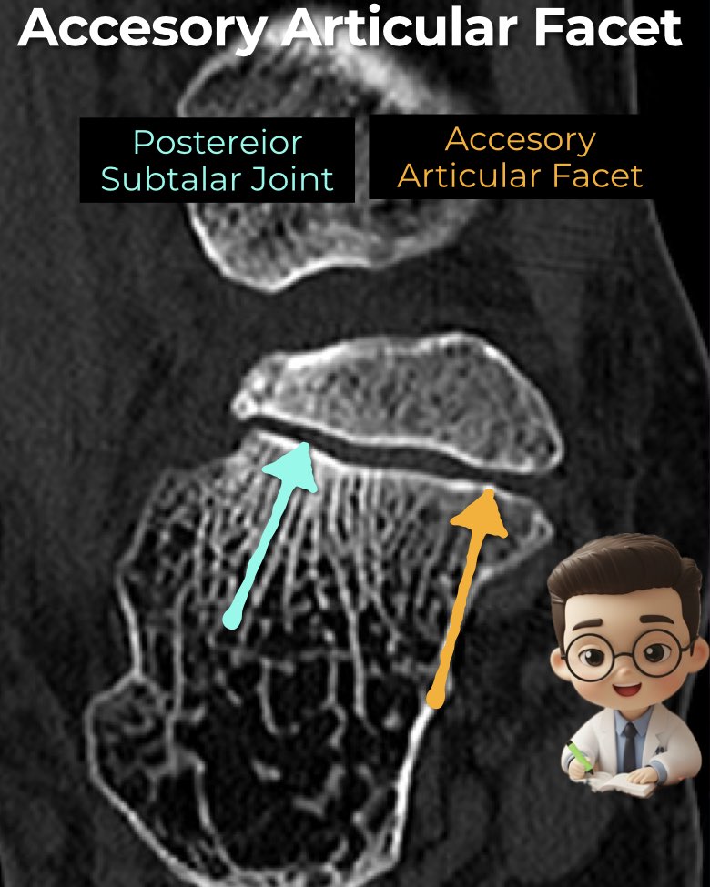
Posteromedial Accessory Facet:
- Located at posterior sustentaculum tali and anteroinferior postero-medial talar process.
- Key point: smooth margins → normal variant, not coalition.
Radiology perspective:
- On MRI, AALTF appears as cartilage extension beyond the lateral talar process, often with adjacent bone marrow edema.
- Recognizing these facets avoids misdiagnosis and helps explain otherwise unexplained subtalar pain.
#Radiology, #MSKMRI, #SubtalarJoint, #AALTF, #FootMRI, #CoalitionImaging, #RadiologyEducation, #OrthopedicImaging, #MRIteaching, #RadiologistLife
Visualizing MSK Radiology: A Practical Guide to Radiology Mastery
© 2022 MSK MRI Jee Eun Lee All Rights Reserved.
No unauthorized reproduction, redistribution, or use for AI training.