https://youtube.com/shorts/7GCBa785WMU
 Coalition by MSKMRI JEE EUN LEE.pdf
6.67MB
Coalition by MSKMRI JEE EUN LEE.pdf
6.67MB
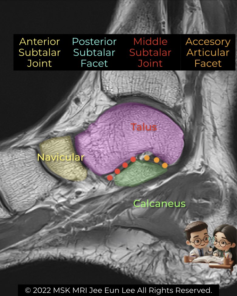
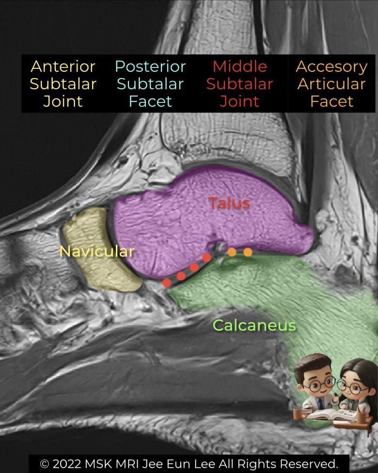
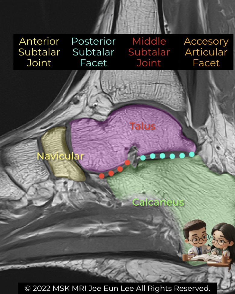
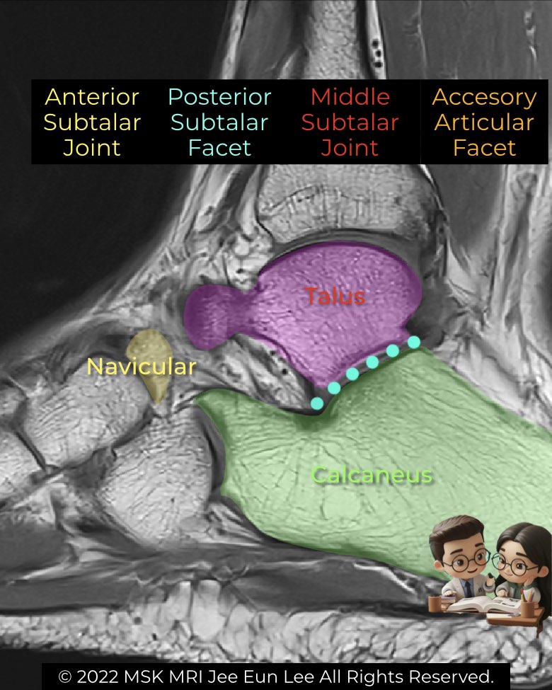
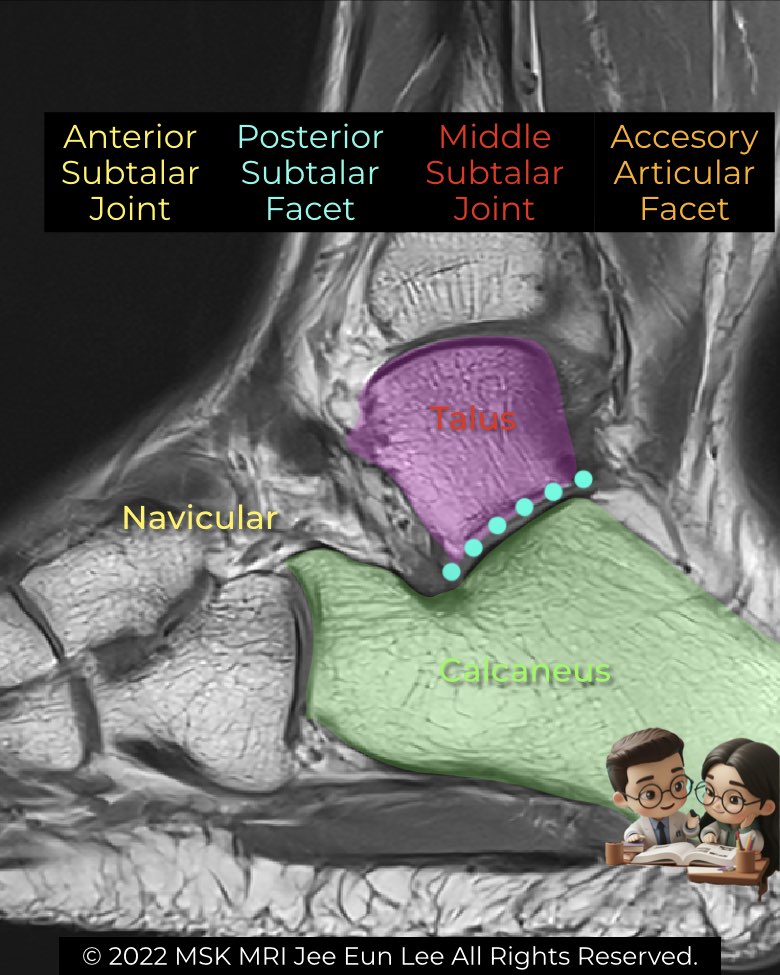
The subtalar joint (talocalcaneal joint) is a key stabilizer of the hindfoot and is well demonstrated on sagittal ankle MRI.
- Posterior facet: the largest articulation, clearly outlined between the talus and calcaneus.
- Middle facet: located on the sustentaculum tali, may show continuity with the talonavicular joint.
- Anterior facet: smallest, sometimes merging with the middle facet.
Imaging pearls (sagittal focus):
- Sustentaculum tali provides a solid platform beneath the talar head, recognizable on sagittal MRI.
- The anterior calcaneal process should remain separate from the navicular—fusion here suggests coalition.
- Harris heel view shows parallel orientation of middle and posterior facets (<20°), but MRI gives superior evaluation of non-osseous variants.
- CT/MRI remain gold standards for characterizing coalition type and subtle accessory anatomy.
#Radiology, #MSKMRI, #SubtalarJoint, #FootMRI, #TalocalcanealJoint, #OrthopedicImaging, #RadiologyEducation, #MRIteaching, #RadiologistLife, #MedicalEducation
Visualizing MSK Radiology: A Practical Guide to Radiology Mastery
© 2022 MSK MRI Jee Eun Lee All Rights Reserved.
No unauthorized reproduction, redistribution, or use for AI training.