https://youtube.com/shorts/BUV5vtYXimk
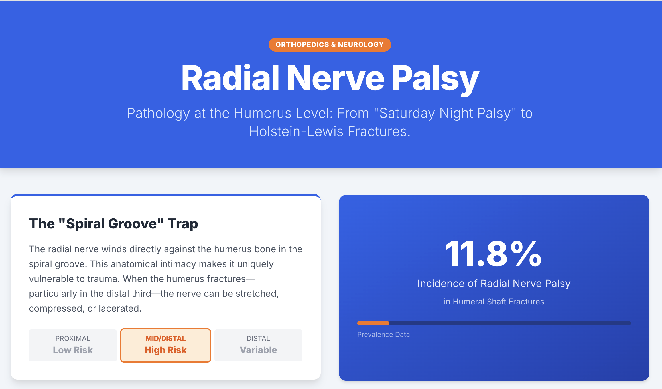
Why the Spiral Groove Matters on MRI
Clinical Context
- Crush injury with compression of the shoulder and upper arm
- Finger extension weakness and arm numbness
- No humeral fracture
Localization
- Finger MCP extension weakness → radial nerve involvement
- Lack of fracture does not rule out nerve injury
Key Anatomy
- Radial nerve courses along the spiral (radial) groove
- Lies directly against the humeral cortex
- Minimal soft-tissue protection → compression-prone
- Distal arm passage through the lateral intermuscular septum = danger zone
MRI Findings (This Case)
- Radial nerve swelling with diffuse T2 hyperintensity
- Extends from spiral groove to distal humerus
- Preserved nerve continuity
- Associated intramuscular hematoma in the brachialis muscle
Interpretation
- Compression- and contusion-related traumatic radial neuropathy
- Injury within the neurapraxia–axonotmesis spectrum
- Important to state: no definite nerve transection
Clinical Pearl
- Saturday night palsy represents the same mechanism
- Prolonged external compression at the spiral groove
- Typically neurapraxia with spontaneous recovery
Take-Home Message
Radial nerve palsy does not require a fracture.
Understanding the anatomy of the spiral groove allows MRI findings, symptoms, and prognosis to align.
#DrSlothic #SlothicRadiology #RadialNerve #RadialNervePalsy #CompressionNeuropathy #SpiralGroove #PeripheralNerveInjury #UpperArmMRI #MusculoskeletalRadiology #NeuroMRI #SaturdayNightPalsy #TraumaticNeuropathy


'✅ Dr. Slothic Notes' 카테고리의 다른 글
| 📌 Unstable OCD in Adults: What MRI Is Really Telling You (0) | 2026.01.18 |
|---|---|
| 📌 The Gravid Hip: Subchondral Insufficiency Fracture of the Femoral Head (Pregnancy & Puerperium) (0) | 2026.01.18 |
| 📌 Aseptic Loosening After Total Knee Arthroplasty (0) | 2026.01.16 |
| 📌 Superior Capsular Reconstruction (SCR): Postoperative MRI (0) | 2026.01.15 |
| 📌 Plantar Fasciitis and Haglund’s Syndrome (0) | 2026.01.14 |