(Fig 1-B.35) Guess what? Classification of Ramp Injuries
https://youtube.com/shorts/ZHs4E3wbEzA?feature=share
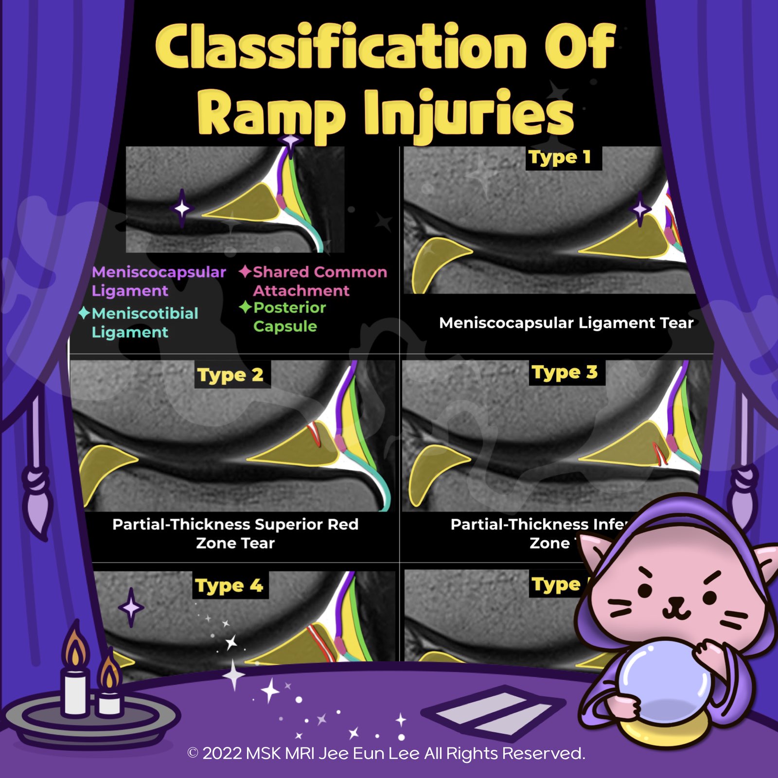
Ramp lesions are categorized into five types based on their characteristics.
Type 1 is an isolated posterior superior meniscocapsular tear, while type 2 involves a partial tear in the same region.
Type 3 represents either a partial posterior inferior tear or a meniscotibial ligament tear.
Type 4 includes a complete posterior peripheral tear or a complete tear at the meniscocapsular junction.
Lastly, type 5 is characterized by a posterior horn double tear.
In terms of stability, ramp lesions type 1 and 2 are generally regarded as stable.
Conversely, types 3, 4, and 5 are considered unstable.
MRI, particularly when interpreted by trained musculoskeletal radiologists, has demonstrated reliable reproducibility in classifying the stability of meniscal ramp lesions, aligning well with arthroscopic assessments.
"Visualizing MSK Radiology: A Practical Guide to Radiology Mastery"
© 2022 MSK MRI Jee Eun Lee All Rights Reserved.
#VisualizingMSK #Ramplesions #ACLinjuries #Medialmeniscus #Meniscaltears
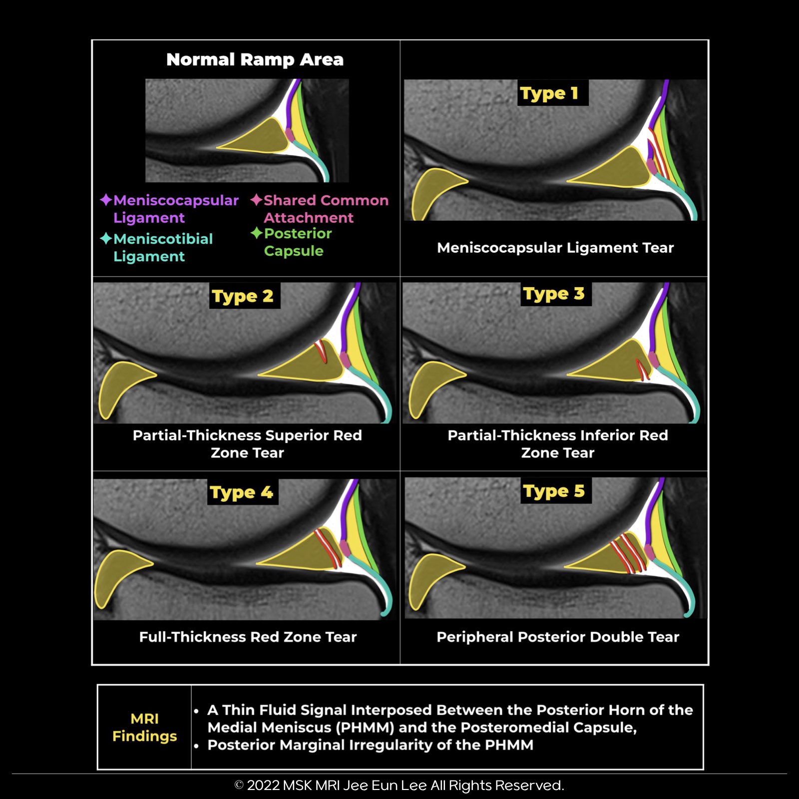
Ramp lesions are categorized into five types based on their characteristics.
- Type 1 is an isolated posterior superior meniscocapsular tear, while type 2 involves a partial tear in the same region.
- Type 3 represents either a partial posterior inferior or meniscotibial ligament tear.
- Type 4 includes a complete posterior peripheral tear or a complete tear at the meniscocapsular junction.
- Lastly, type 5 is characterized by a posterior horn double tear.
In terms of stability, ramp lesions type 1 and 2 are generally regarded as stable.
Conversely, types 3, 4, and 5 are considered unstable.
MRI, particularly when interpreted by trained musculoskeletal radiologists, has demonstrated reliable reproducibility in classifying the stability of meniscal ramp lesions, aligning well with arthroscopic assessments.
| MRI Features of Ramp Leions | |
| 1 | A thin fluid signal completely interposed between the posterior horn of the medial meniscus and the posteromedial capsule. |
| 2 | Longitudinal vertical and/or oblique tear affecting the peripheral zone of the posterior horn of the medial meniscus. |
| 3 | Irregularities involving the posterior margin of medial meniscus, with focal discontinuity or step-like deformity, and involving capsular attachments. |
| 4 | Soft tissue edema between meniscus and collateral ligament. |
| 5 | Bone bruise of posteromedial tibia from pivot shift countercoup injury in medial compartment, and anterior translation of medial plateau in relation to femoral condyle. |
| 6 | Signs of concurrent ACL injury. |
"Visualizing MSK Radiology: A Practical Guide to Radiology Mastery"
© 2022 MSK MRI Jee Eun Lee All Rights Reserved.
#VisualizingMSK #Ramplesions #ACLinjuries #Medialmeniscus #Meniscaltears