https://youtube.com/shorts/oZnueY6FxO8
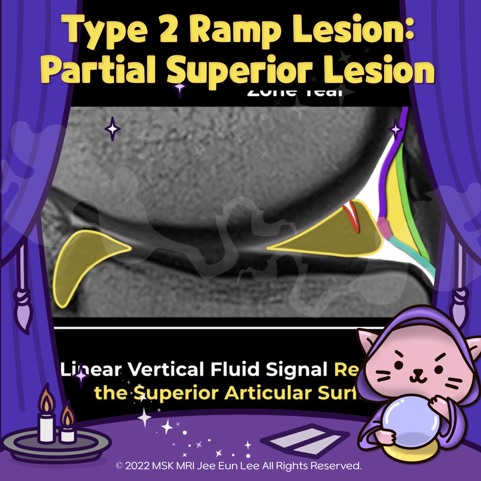
Type 2 Ramp Lesion: Partial Superior Lesion
- Type 2 Ramp Lesions are characterized by peripheral partial-thickness tears involving the superior margin of the posterior horn of the medial meniscus.
- These tears retain intact meniscocapsular attachments to the posterior horn.
- This type of tear pattern is generally considered more stable compared to full-thickness meniscocapsular tears.
MRI Findings:
- The image depicts a Type 2 Ramp Lesion, showing a partial thickness superior vertical tear extending through the posterior horn of the medial meniscus, with a linear vertical fluid signal reaching the superior articular surface.
- Additionally, a peripheral partial tear of the posterior horn of the medial meniscus affects the femoral articular surface, accompanied by a meniscocapsular ligament tear.
- Bone Marrow Edema:
This is present in the anterior aspect of the medial femoral condyle and posterior margin of the medial tibial plateau, indicative of recent contrecoup injury.
"Visualizing MSK Radiology: A Practical Guide to Radiology Mastery"
© 2022 MSK MRI Jee Eun Lee All Rights Reserved.
#VisualizingMSK #Ramplesions #ACLinjuries #Medialmeniscus #Meniscaltears


'✅ Knee MRI Mastery > Chap 1. Meniscus' 카테고리의 다른 글
| (Fig 1-B.39) Type 4A Ramp lesion (0) | 2024.02.07 |
|---|---|
| (Fig 1-B.38) Type 3 Ramp lesion, Partial inferior or hidden lesion (0) | 2024.02.07 |
| (Fig 1-B.36) Type 1 Ramp lesion, Meniscocapsular lesion (0) | 2024.02.07 |
| (Fig 1-B.35) Classification of Ramp Injuries (0) | 2024.02.06 |
| (Fig 1-B.34) Type 4 complex oblique or longitudinal tear with complete root detachment (0) | 2024.02.06 |