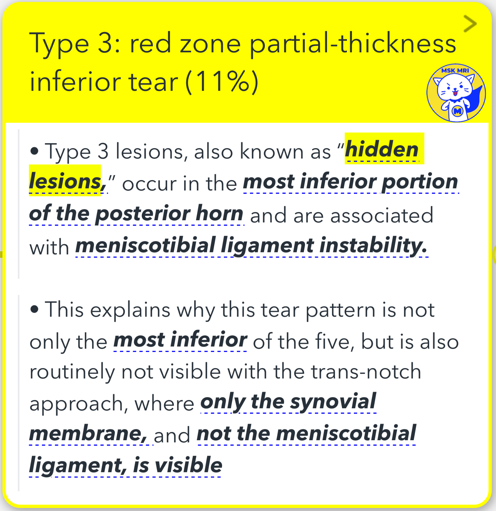
1. Type 3 Ramp Lesions - 'Hidden Lesions':
- Occur in the most inferior portion of the posterior horn.
- Associated with meniscotibial ligament instability.
- Characterized by a red zone partial-thickness inferior tear, accounting for about 11% of cases.
- Not typically visible with the trans-notch approach due to their location and partial nature, making them easy to miss on preoperative MRI scans.
- Diagnostic indicators on MRI include a thin fluid signal between the posterior horn of the medial meniscus and the posteromedial capsule and posterior marginal irregularity.
2. Type 3A Lesion:
- Defined as a vertical peripheral tear at the inferior margin of the posterior horn.
- Involves the attachment of the meniscotibial ligament.
- MRI findings may show linear vertical oblique fluid intensity signal reaching the inferior articular surface and discontinuity of the meniscotibial ligament with the meniscotibial ligament still attached to the torn meniscus
- Sometimes, it presents as a partial separation of the meniscocapsular junction, leading to a corner lesion.
3. Type 3B Lesion:
- Characterized by a partial inferior tear affecting the meniscotibial ligament.
- Represents a tear of the meniscotibial ligament itself from its attachment to the posterior horn.
- Includes rupture in the mid-substance of the ligament or an avulsion from the meniscal insertion.
- MRI may show disruption of the ligament with high T2 signal and possibly associated bone marrow edema pattern within the posterior margin of the medial tibial plateau from recent contrecoup injury."
"Visualizing MSK Radiology: A Practical Guide to Radiology Mastery"
© 2022 MSK MRI Jee Eun Lee All Rights Reserved.
#VisualizingMSK #Ramplesions #ACLinjuries #Hiddenlesion


'✅ Knee MRI Mastery > Chap 1. Meniscus' 카테고리의 다른 글
| (Fig 1-B.40) Type 4B Ramp lesion (0) | 2024.02.07 |
|---|---|
| (Fig 1-B.39) Type 4A Ramp lesion (0) | 2024.02.07 |
| (Fig 1-B.37) Type 2 Ramp lesion, Partial superior lesion (0) | 2024.02.07 |
| (Fig 1-B.36) Type 1 Ramp lesion, Meniscocapsular lesion (0) | 2024.02.07 |
| (Fig 1-B.35) Classification of Ramp Injuries (0) | 2024.02.06 |