Key points in identifying and assessing a meniscal flounce include:
- ✅Appearance:
It presents as a wavy or kinked pattern along the meniscus's inner edge. - ✅Imaging Challenge:
On coronal plane imaging, a flounce can make the meniscus's inner margin appear shortened or fuzzy, which might be mistaken for a small radial tear. - ✅Clinical Significance:
The identification of a flounce necessitates a thorough check for any associated ligamentous or capsular injuries, as these could contribute to the laxity seen in the meniscus. - ✅Special Consideration for Lateral Meniscal Flounce: Flounces in the lateral meniscus are uncommon.
Their presence could indicate an injury or deficiency in the popliteomeniscal fascicles, leading to a hypermobile meniscus requiring careful evaluation.
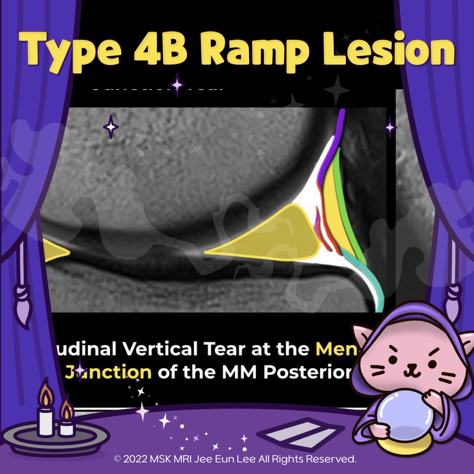
Subtype 4B:
❗️This pattern involves a complete tear at the junction where the meniscocapsular and meniscotibial fibers attach to the posterior horn of the meniscus.
❗️MRI reveals a linear high T2 fluid signal intensity extending from the superior to the inferior articular surface, along with disruption of the capsular ligaments.
❗️The junction consists only of meniscocapsular and meniscotibial fibers. As a result, it does not share the same healing capacity as lesions within the red-red zone of the meniscus, often requiring more extensive repair measures.
"Visualizing MSK Radiology: A Practical Guide to Radiology Mastery"
© 2022 MSK MRI Jee Eun Lee All Rights Reserved.
#VisualizingMSK #Ramplesions #ACLinjuries #meniscocapsularseparation


'✅ Knee MRI Mastery > Chap 1. Meniscus' 카테고리의 다른 글
| (Fig 1-B.43) Wrisberg rip tear (0) | 2024.02.07 |
|---|---|
| (Fig 1-B.41) False positive Ramp lesions (0) | 2024.02.07 |
| (Fig 1-B.39) Type 4A Ramp lesion (0) | 2024.02.07 |
| (Fig 1-B.38) Type 3 Ramp lesion, Partial inferior or hidden lesion (0) | 2024.02.07 |
| (Fig 1-B.37) Type 2 Ramp lesion, Partial superior lesion (0) | 2024.02.07 |