https://youtube.com/shorts/Kno6P7UGi4E?si=8WqESkAXGd0r0fbQ
📊Essentials of the Wrisberg Rip and Meniscofemoral Ligaments 📊
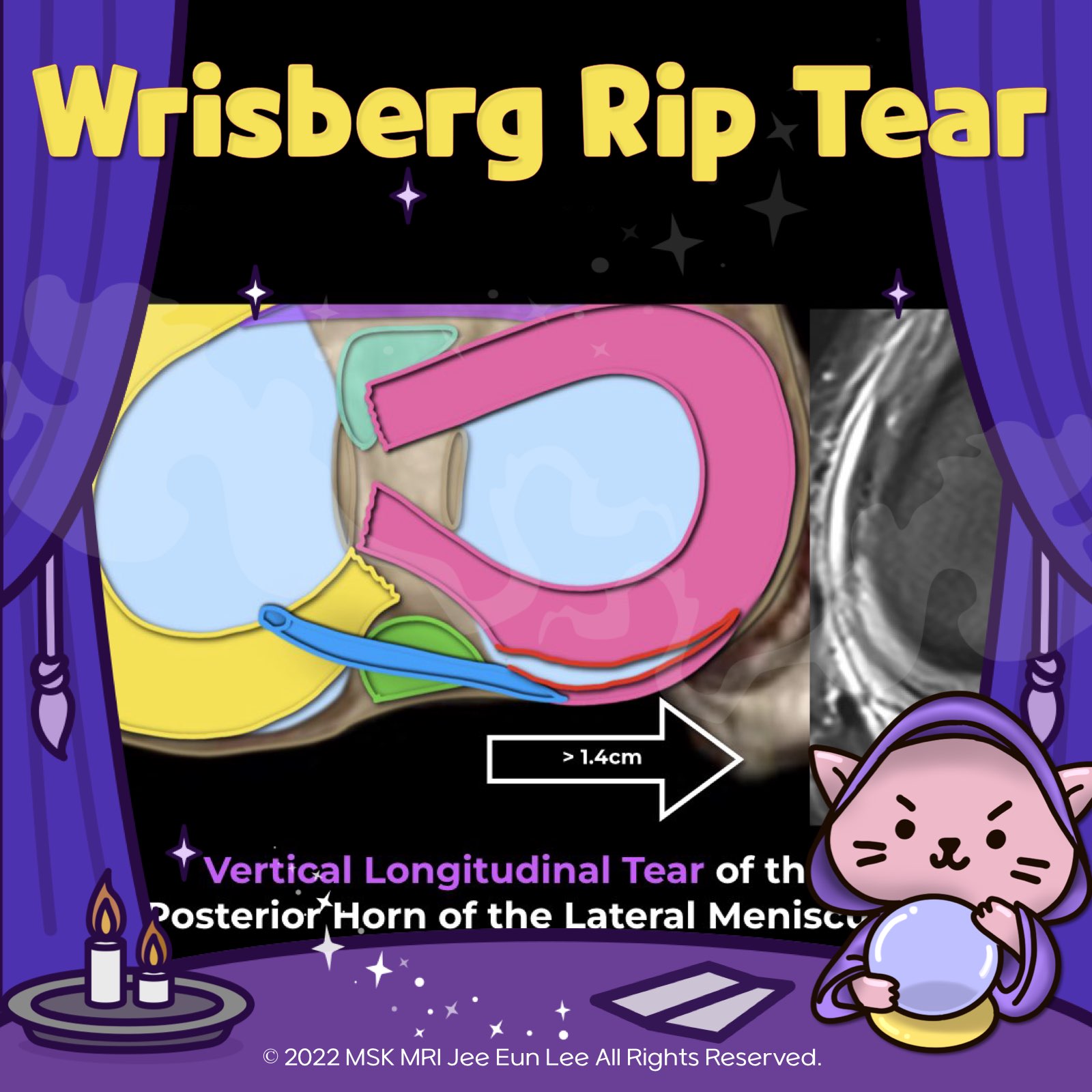
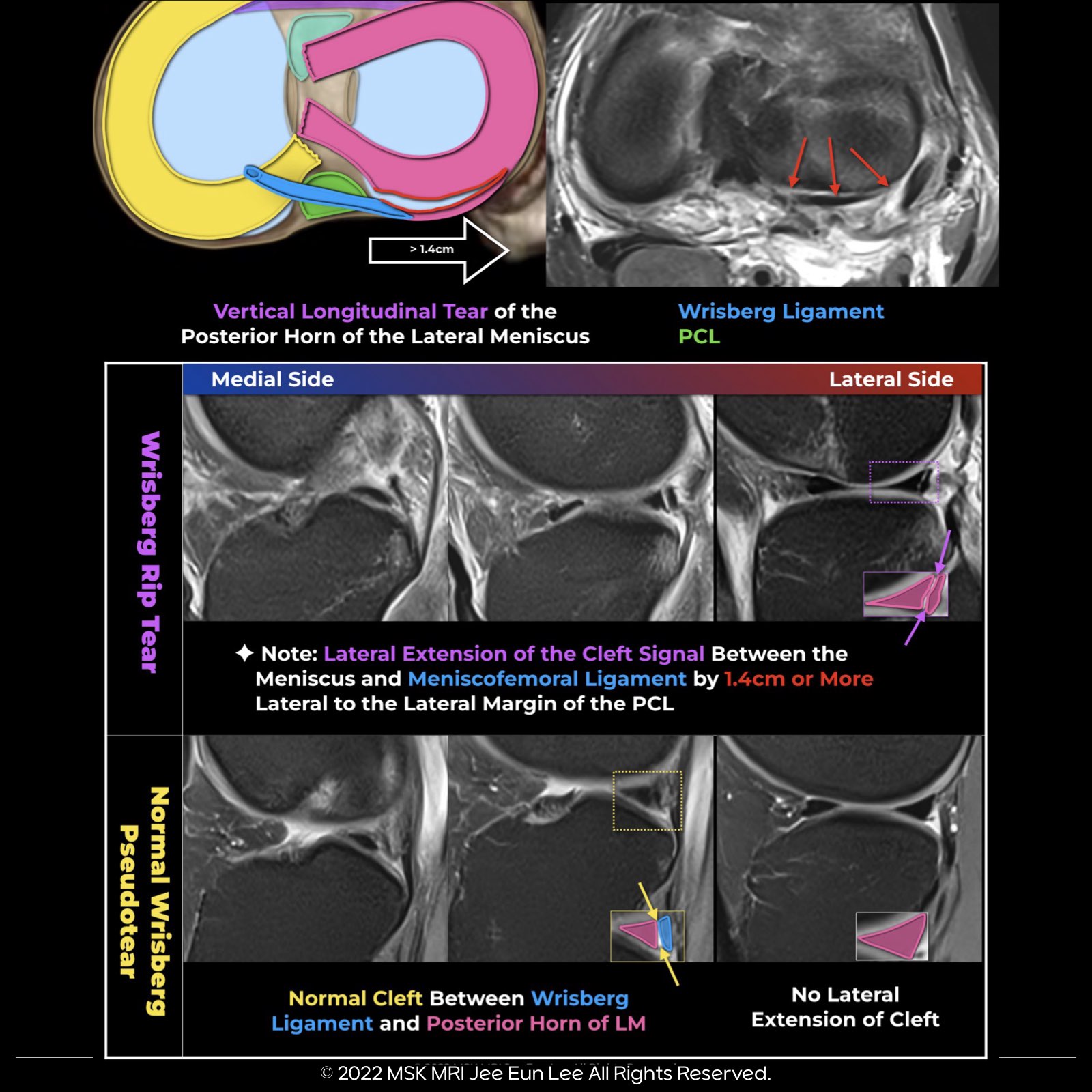
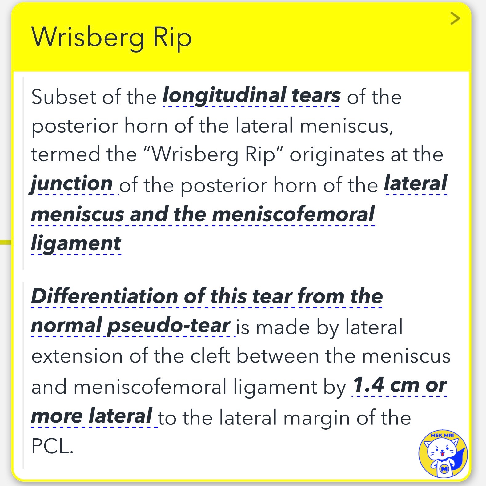
1. Wrisberg Rip 🌟:
- Identifies a specific longitudinal tear where the posterior horn of the lateral meniscus meets the meniscofemoral ligament.
- Look for this if the meniscofemoral ligament extends 14 mm past the PCL — a key sign of a peripheral longitudinal tear.
2. Meniscofemoral Ligament (MFL) 💡:
- Starting from the posterior horn of the lateral meniscus, this ligament attaches to the posterior medial femoral condyle. It's divided into the anterior Humphry ligament and the posterior Wrisberg ligament.
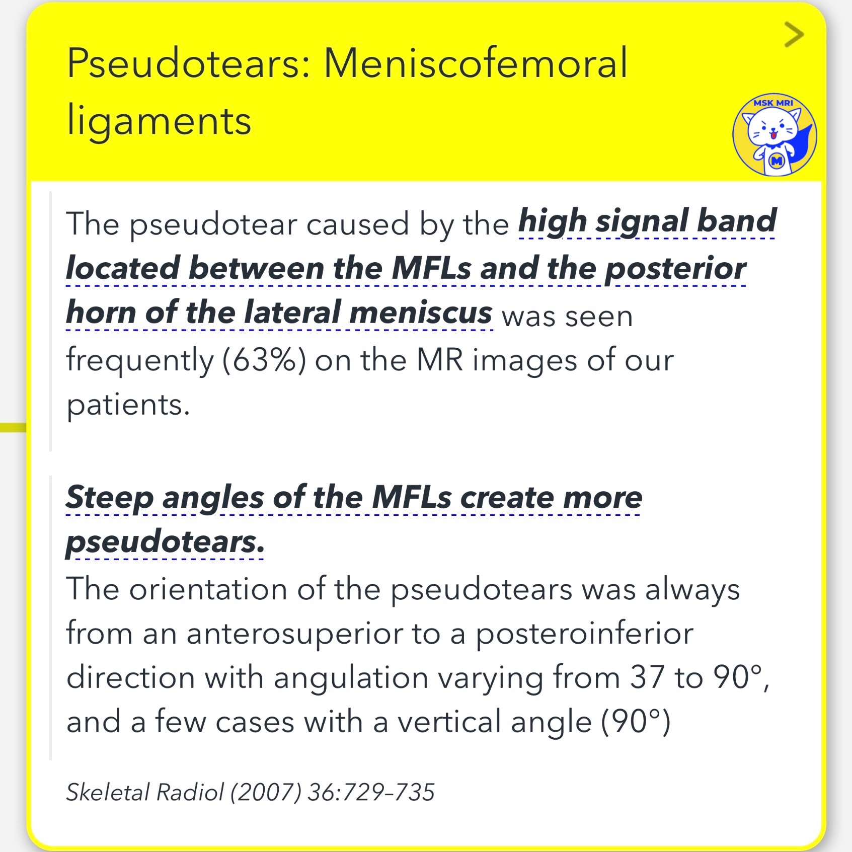
3. Wrisberg Pseudotears 🔍:
- Common in MRIs (seen in about 63% of cases), these pseudotears between the MFLs and the posterior horn of the lateral meniscus can be easily confused with actual peripheral tears, especially Wrisberg Rip.
"Visualizing MSK Radiology: A Practical Guide to Radiology Mastery"
© 2022 MSK MRI Jee Eun Lee All Rights Reserved.
#VisualizingMSK #WrisbergRip #MeniscofemoralLigament #LateralMeniscus #Pseudotears #ACLinjuries #SportsInjuryMRI
'✅ Knee MRI Mastery > Chap 1. Meniscus' 카테고리의 다른 글
| (Fig 1-B.45) Radial tear sparing the MFL attachment without displacement (0) | 2024.02.07 |
|---|---|
| (Fig 1-B.44) Complete complex tear, including MFL attachment (0) | 2024.02.07 |
| (Fig 1-B.41) False positive Ramp lesions (0) | 2024.02.07 |
| (Fig 1-B.40) Type 4B Ramp lesion (0) | 2024.02.07 |
| (Fig 1-B.39) Type 4A Ramp lesion (0) | 2024.02.07 |