✅ Meniscal Flounce Summary:
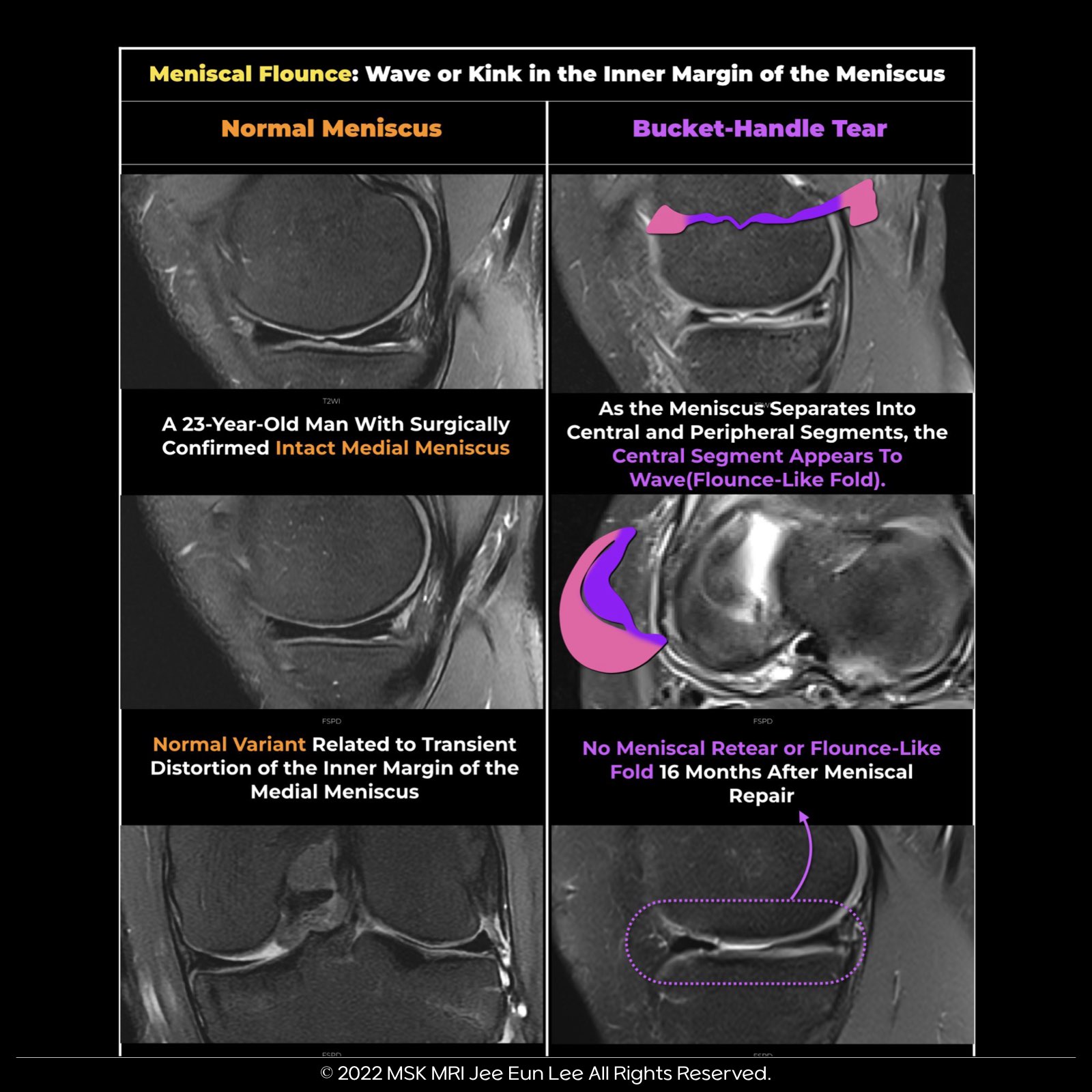
- Meniscal flounce refers to a wave-like appearance of meniscus edges in sagittal MRI images.
- It occurs in about 0.2% to 0.3% of patients, mainly when the knee is flexed, and the free edge shows redundancy.
- While discussed in the context of the medial meniscus, it can also affect the lateral meniscus.
- The wave-like appearance is notable, but other attachments are usually intact, and no tearing is seen in coronal images.
🧐 Key Points to Consider:
✅ Pitfall:
- Flounce may mimic a torn meniscus in sagittal images but is not indicative of an actual tear.
- Coronal images can make the free edge look torn due to the wave-like shape.
✅ Don't Dismiss Flounce:
- When a meniscal flounce is detected, it's crucial to examine for potential ligamentous or capsular injuries that might cause laxity.
- Detecting a meniscal flounce necessitates checking for possible ligamentous or capsular injuries causing laxity, particularly with lateral meniscal flounces.
- These are rare and might suggest popliteomeniscal fascicle injury/deficiency, resulting in meniscal hypermobility.
"Visualizing MSK Radiology: A Practical Guide to Radiology Mastery"
© 2022 MSK MRI Jee Eun Lee All Rights Reserved.
#VisualizingMSK #meniscaltear #Flounce #KneeMRI #Meniscus


'✅ Knee MRI Mastery > Chap 1. Meniscus' 카테고리의 다른 글
| (Fig 1-C.18) Meniscal Ossicle (1) | 2024.02.09 |
|---|---|
| (Fig 1-C.17) Meniscal Flounce with meniscocapsular tear (0) | 2024.02.08 |
| (Fig 1-C.15) Lateral Meniscal Extrusion (0) | 2024.02.08 |
| (Fig 1-C.14) Medial Meniscal Extrusion (0) | 2024.02.08 |
| (Fig 1-C.13) Parameniscal cyst versus ganglion cysts (0) | 2024.02.08 |