Click the link to purchase on Amazon 🎉📚
==============================================
🎥 Check Out All Videos at Once! 📺
👉 Visit Visualizing MSK Blog to explore a wide range of videos! 🩻
https://visualizingmsk.blogspot.com/?view=magazine
📚 You can also find them on MSK MRI Blog and Naver Blog! 📖
https://www.instagram.com/msk_mri/
Click now to stay updated with the latest content! 🔍✨
==============================================
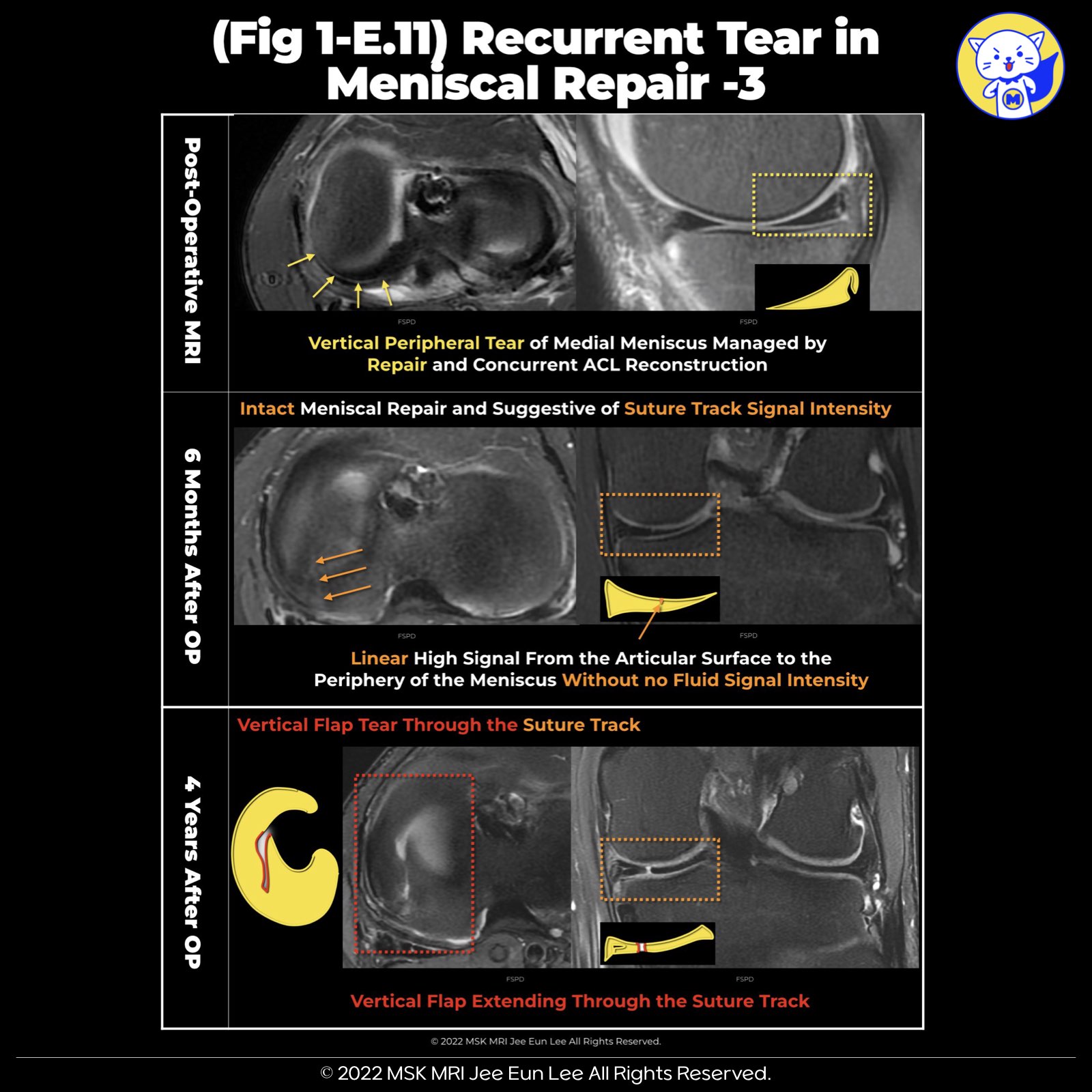
📌 Suture Tracks in Meniscal Tissue
Identification: Suture tracks within the meniscal tissue are discernible across all MRI pulse sequences.
However, their visibility is notably enhanced in PD (Proton Density) Fat Suppressed (PDFS) sequences.
➡️ Suture Tracks Appearance:
- Short Axis: Manifest as dot-like images, showcasing an intermediate to high signal.
- Long Axis: Present as linear images, also displaying an intermediate to high signal.
➡️ Suture Tracks Location:
- These suture tracks are embedded within the thickness of the meniscal tissue.
- A detailed examination allows for their tracing from the articular surface to the meniscus's periphery.
📌 Radial Tears in Vertical Suture
- It is possible to observe their formation originating from the suture tracks, extending towards the femoral and tibial surfaces of the meniscus
"Visualizing MSK Radiology: A Practical Guide to Radiology Mastery"
© 2022 MSK MRI Jee Eun Lee All Rights Reserved.
#VisualizingMSK #meniscaltear #meniscus #meniscectomy #meniscalrepair #meniscalretear
You may not distribute or commercially exploit the content.
Nor may you transmit it or store it on any other website or other forms of the electronic retrieval system.


'✅ Knee MRI Mastery > Chap 1. Meniscus' 카테고리의 다른 글
| (Fig 1-E.13) Transtibial Pullout Repair (0) | 2024.02.10 |
|---|---|
| (Fig 1-E.12) Recurrent Tear in meniscal repair -4 (2) | 2024.02.10 |
| (Fig 1-E.10) Recurrent Tear in meniscal repair -2 (0) | 2024.02.10 |
| (Fig 1-E.09) Recurrent Tear in meniscal repair -1 (0) | 2024.02.10 |
| (Fig 1-E.08) Expected appearance of meniscal repair -3 (0) | 2024.02.09 |