Click the link to purchase on Amazon 🎉📚
==============================================
🎥 Check Out All Videos at Once! 📺
👉 Visit Visualizing MSK Blog to explore a wide range of videos! 🩻
https://visualizingmsk.blogspot.com/?view=magazine
📚 You can also find them on MSK MRI Blog and Naver Blog! 📖
https://www.instagram.com/msk_mri/
Click now to stay updated with the latest content! 🔍✨
==============================================
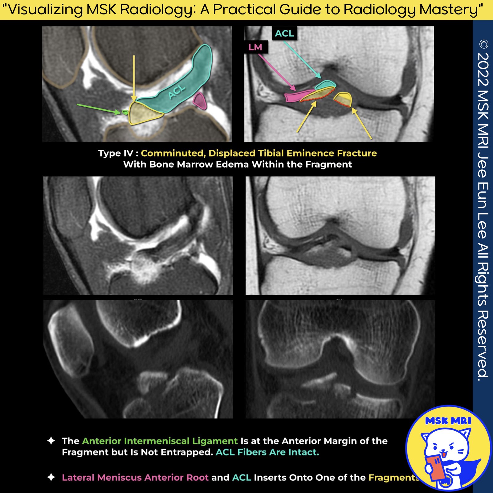
✅ Meyers and McKeever system (with modifications by Zaricznyj)
Type 1: Minimally/Nondisplaced Fragment
Type 2: Anterior Elevation Of The Fragment
Type 3: Complete Separation Of The Fragment
Type 3a: Involves A Small Portion Of Eminence
Type 3b: Involves The Majority Of The Eminence
Type 4: Comminuted Avulsion Or Rotation Of The Fracture Fragment
1. Lateral Meniscus and ACL Attachments:
The study highlights a significant overlap (approximately 40%) between the anterior root of the lateral meniscus and the ACL's tibial attachment, observed under an electron microscope.
Notably, avulsed fracture fragments adhered to the anterior horn of the lateral meniscus.
2. CT Scans versus FSPD in Bone Fragment Visibility:
CT scans are superior in visualizing bone fragments compared to FSPD, mainly due to the clarity provided by bone marrow edema, surrounding fluid, and hemorrhage.
"Visualizing MSK Radiology: A Practical Guide to Radiology Mastery"
© 2022 MSK MRI Jee Eun Lee All Rights Reserved.
#VisualizingMSK #ACLinjuries #KneeMRI #ACLtear #ACLavulsion #Avulsionfracture
You may not distribute or commercially exploit the content.
Nor may you transmit it or store it on any other website or other forms of the electronic retrieval system.


'✅ Knee MRI Mastery > Chap 2.ACL and PCL' 카테고리의 다른 글
| (Fig 2-B.19) Low-grade Partial tears of the ACL (1) | 2024.02.23 |
|---|---|
| (Fig 2-B.18) Meyers and McKeever classification system type IV (0) | 2024.02.23 |
| (Fig 2-B.16) Meyers and McKeever classification system type II (0) | 2024.02.22 |
| (Fig 2-B.15) ACL Avulsion fracture from the femoral attachment (0) | 2024.02.21 |
| (Fig 2-B.14) Type 2 ACL stump entrapment Lesion (0) | 2024.02.21 |