Click the link to purchase on Amazon 🎉📚
==============================================
🎥 Check Out All Videos at Once! 📺
👉 Visit Visualizing MSK Blog to explore a wide range of videos! 🩻
https://visualizingmsk.blogspot.com/?view=magazine
📚 You can also find them on MSK MRI Blog and Naver Blog! 📖
https://www.instagram.com/msk_mri/
Click now to stay updated with the latest content! 🔍✨
==============================================
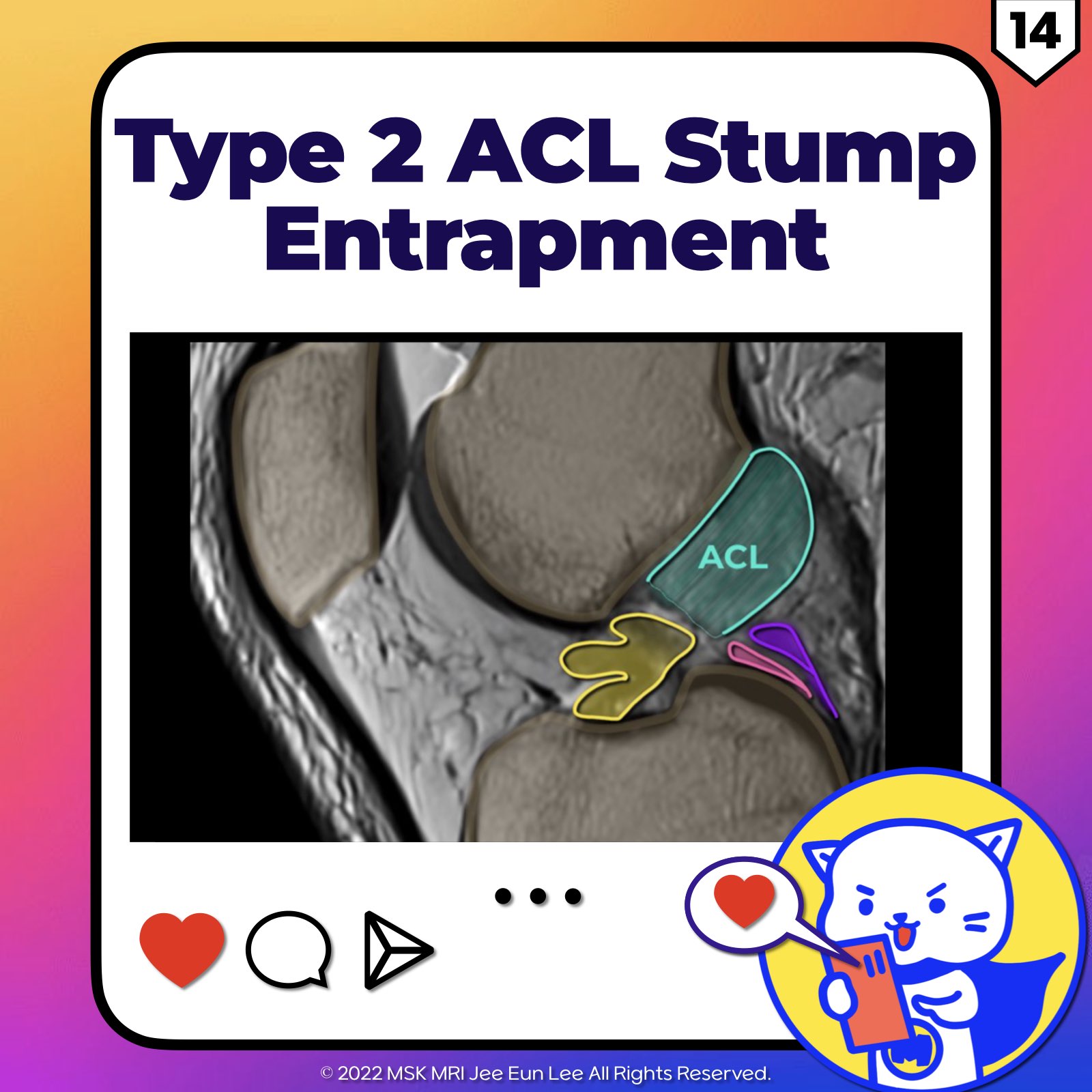
✅Stump Entrapment (SE) Lesions in ACL Injuries ✅
Stump entrapment (SE) lesions are critical considerations in the assessment of anterior cruciate ligament (ACL) injuries, presenting in two distinct types, each with unique morphological characteristics.
1️⃣ Type 1 SE Lesions:
- Characteristics and Formation Type 1 SE lesions typically mimic the appearance of a classic post-operative cyclops lesion characterized by a nodular mass.
- These lesions are predominantly located at the anterior aspect of the intercondylar notch.
- The formation of Type 1 SE lesions is believed to evolve from Type 2 SE lesions, as proposed by Huang et al.
- type 1 SE lesion develops from a type 2 SE lesion, due to chronic impingement of the ACL stump between femur and tibia, resulting in fibrotic changes
- This transition is attributed to the chronic impingement of the ACL stump between the femur and tibia, leading to fibrotic changes.
2️⃣ Type 2 SE Lesions:
- Description In contrast, Type 2 SE lesions exhibit a distinctive morphology, lacking the mass-like appearance characteristic of Type 1 lesions.
- Instead, they feature the anterior portion of the torn ACL folded upon itself, resulting in a thin, tongue-like free end that extends anteriorly out of the intercondylar notch.
"Visualizing MSK Radiology: A Practical Guide to Radiology Mastery"
© 2022 MSK MRI Jee Eun Lee All Rights Reserved.
#VisualizingMSK #ACLinjuries #KneeMRI #ACLtear #Stumpentrapment #ACLstump
You may not distribute or commercially exploit the content.
Nor may you transmit it or store it on any other website or other forms of the electronic retrieval system.


'✅ Knee MRI Mastery > Chap 2.ACL and PCL' 카테고리의 다른 글
| (Fig 2-B.16) Meyers and McKeever classification system type II (0) | 2024.02.22 |
|---|---|
| (Fig 2-B.15) ACL Avulsion fracture from the femoral attachment (0) | 2024.02.21 |
| (Fig 2-B.13) Type 1 ACL stump entrapment Lesion (0) | 2024.02.21 |
| (Fig 2-B.12) Positive PCL angle and sign (1) | 2024.02.21 |
| (Fig 2-B.11) Secondary Signs of the Acute ACL Tears, Anterior Tibial Translocation (0) | 2024.02.20 |