Click the link to purchase on Amazon 🎉📚
==============================================
🎥 Check Out All Videos at Once! 📺
👉 Visit Visualizing MSK Blog to explore a wide range of videos! 🩻
https://visualizingmsk.blogspot.com/?view=magazine
📚 You can also find them on MSK MRI Blog and Naver Blog! 📖
https://www.instagram.com/msk_mri/
Click now to stay updated with the latest content! 🔍✨
==============================================
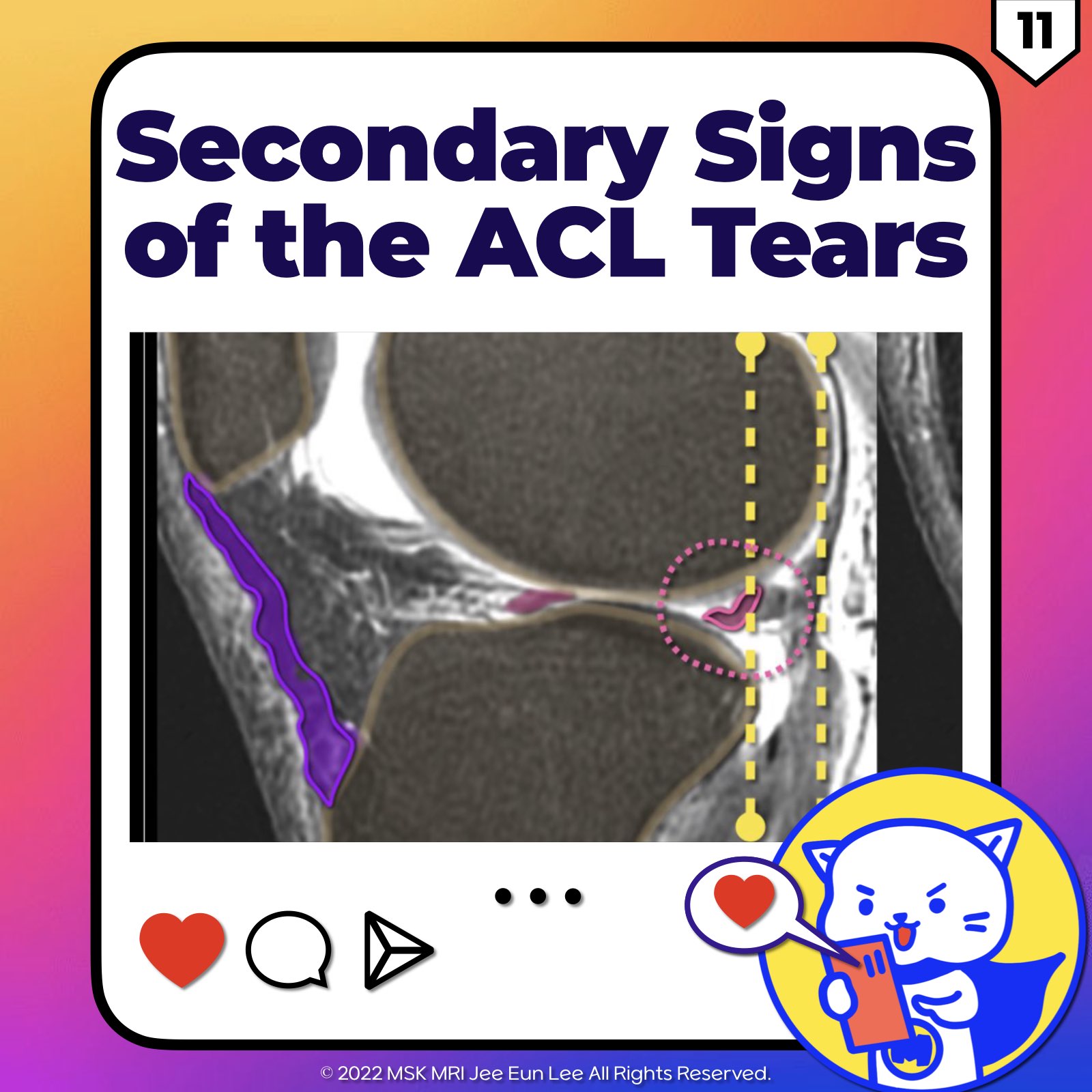
✅ Secondary Signs of ACL Injury
Secondary signs are highly specific indicators of an Anterior Cruciate Ligament (ACL) injury, though they are less sensitive compared to primary signs.
While primary signs are pivotal for assessing the ACL's integrity, secondary signs add limited extra diagnostic value. Here's a brief overview:
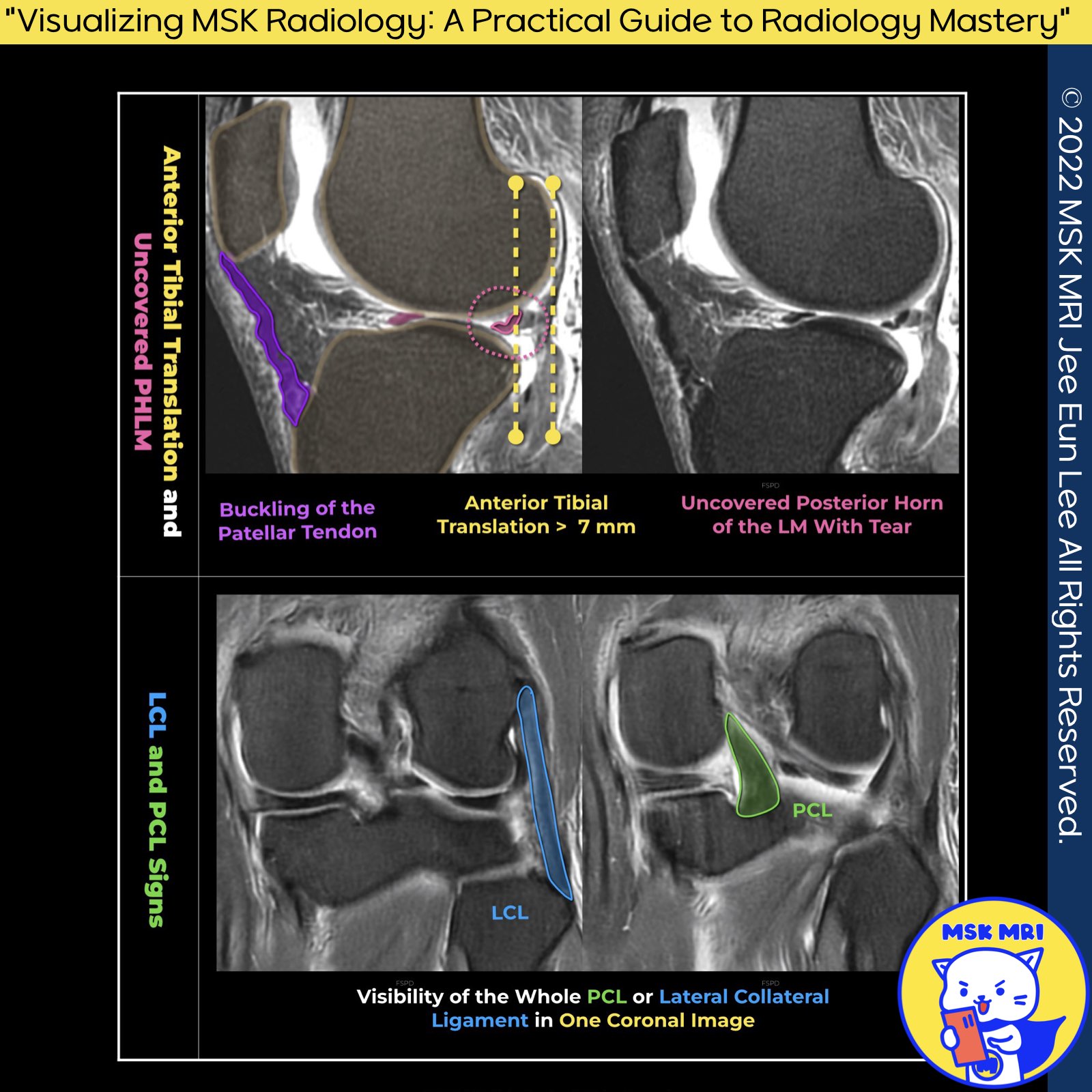
1️⃣ Anterior Translation of the Tibia (Anterior Tibial Translocation):
- The ACL prevents anterior movement of the tibia. In cases of ACL tears, increased anterior tibial translation, known as "anterior tibial translocation," is observed during knee extension.
- In complete ACL ruptures, this sign involves measuring anterior tibial translation relative to the lateral femoral condyle in the mid-sagittal plane.
- Translation between 5 and 7 mm is suggestive, while more than 7 mm confirms an ACL tear. This assessment also reveals potential lateral meniscus (LM) tears.
2️⃣ Uncovering of the Posterior Horn of the Lateral Meniscus:
- This sign indicates a posterior displacement of the lateral meniscus's posterior horn relative to the lateral tibial plateau's posterior aspect, hinting at an ACL injury.
3️⃣ Coronal Whole PCL and LCL Sign:
- The visibility of the popliteus tendon, Lateral Collateral Ligament (LCL), and biceps femoris on a single coronal image suggests an ACL tear.
- An acute ACL disruption causes anterior tibial translation, altering the LCL's orientation to a more coronal position.
"Visualizing MSK Radiology: A Practical Guide to Radiology Mastery"
© 2022 MSK MRI Jee Eun Lee All Rights Reserved.
#VisualizingMSK #ACLinjuries #KneeMRI #ACLtear #fracture #anteriordraw
You may not distribute or commercially exploit the content.
Nor may you transmit it or store it on any other website or other forms of the electronic retrieval system.


'✅ Knee MRI Mastery > Chap 2.ACL and PCL' 카테고리의 다른 글
| (Fig 2-B.13) Type 1 ACL stump entrapment Lesion (0) | 2024.02.21 |
|---|---|
| (Fig 2-B.12) Positive PCL angle and sign (1) | 2024.02.21 |
| (Fig 2-B.10) Deep notch and Long notch signs (1) | 2024.02.20 |
| (Fig 2-B.09) Mass-like tissue without normally oriented ACL fibers (0) | 2024.02.20 |
| (Fig 2-B.08) Abnormal orientation or bowing of the ACL (0) | 2024.02.20 |