==============================================
⬇️✨⬇️🎉⬇️🔥⬇️📚⬇️
Click the link to purchase on Amazon 🎉📚
==============================================
🎥 Check Out All Videos at Once! 📺
👉 Visit Visualizing MSK Blog to explore a wide range of videos! 🩻
https://visualizingmsk.blogspot.com/?view=magazine
📚 You can also find them on MSK MRI Blog and Naver Blog! 📖
https://www.instagram.com/msk_mri/
Click now to stay updated with the latest content! 🔍✨
==============================================
1️⃣ MRI Imaging Findings for ACL Tears:
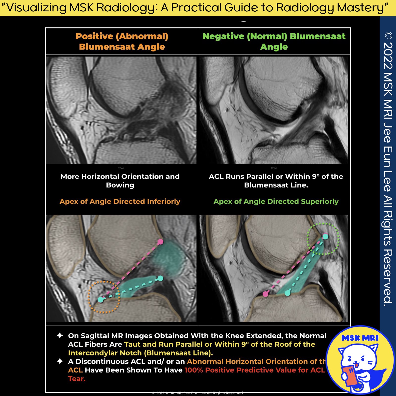
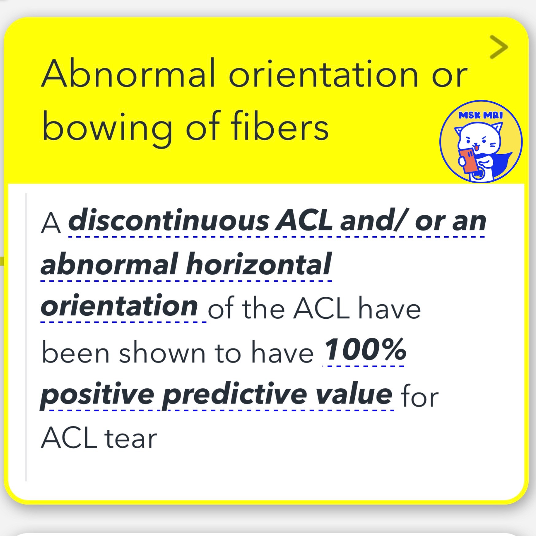
- Positive Predictive Value: A discontinuous anterior cruciate ligament (ACL) or an abnormal horizontal orientation of the ACL is indicative of a 100% positive predictive value for an ACL tear.
2️⃣ Horizontal orientation of ACL:
- An angle greater than 15º between the roof of the intercondylar notch and the ACL suggests a complete tear.
- An angle less than 45º between the distal ACL and the tibia, indicating a horizontal orientation of ACL fibers, is also highly accurate for diagnosing complete ACL tears.
3️⃣ Blumensaat Angle:
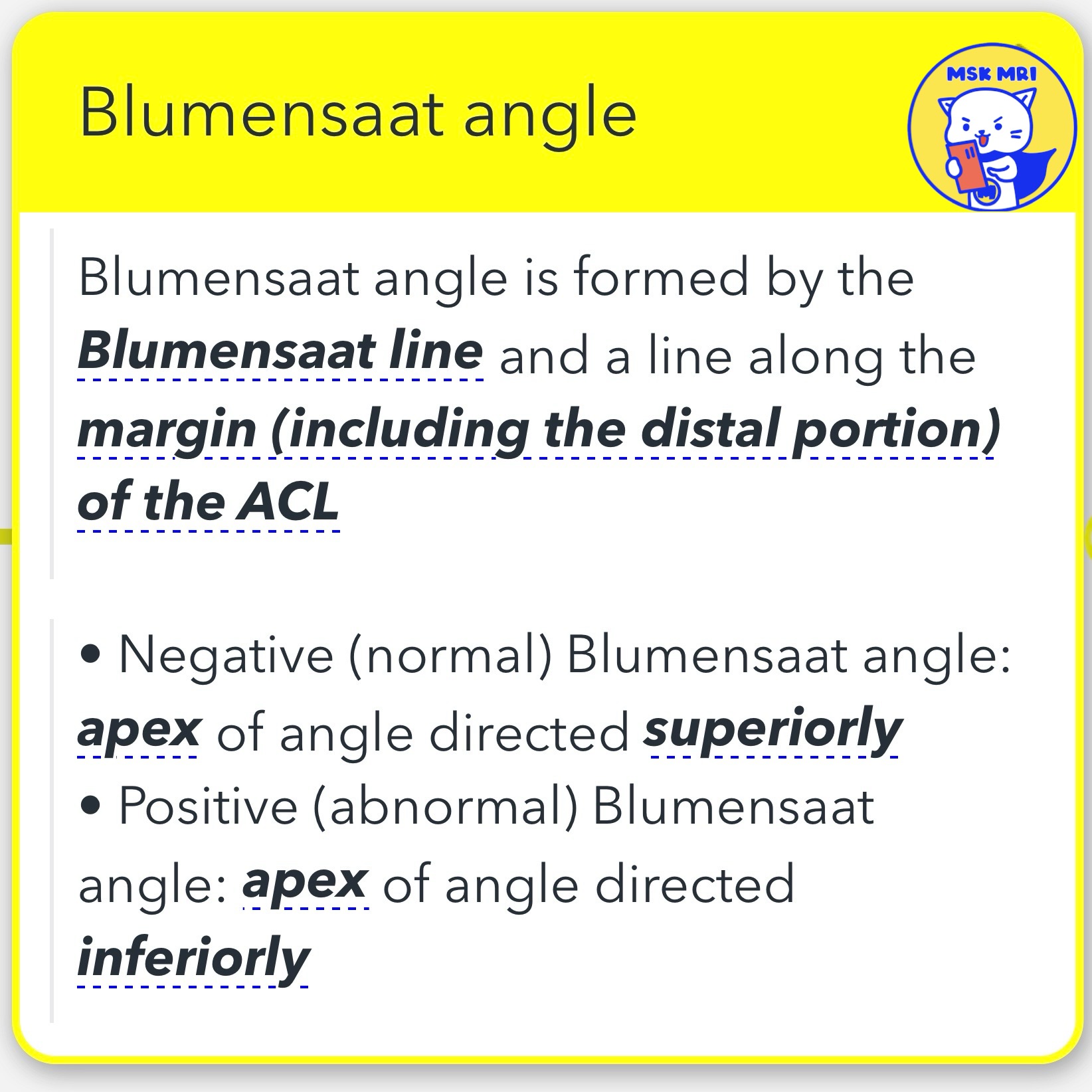
- The Blumensaat angle is created by the intersection of the Blumensaat line and a line along the margin of the ACL, including its distal portion.
- Negative (Normal) Blumensaat Angle: The apex of the angle is directed superiorly.
- Positive (Abnormal) Blumensaat Angle: The apex of the angle is directed inferiorly.
"Visualizing MSK Radiology: A Practical Guide to Radiology Mastery" © 2022 MSK MRI Jee Eun Lee All Rights Reserved.
#VisualizingMSK #ACLinjuries #KneeMRI #ACLtear You may not distribute or commercially exploit the content. Nor may you transmit it or store it on any other website or other forms of the electronic retrieval system.
'✅ Knee MRI Mastery > Chap 2.ACL and PCL' 카테고리의 다른 글
| (Fig 2-B.10) Deep notch and Long notch signs (1) | 2024.02.20 |
|---|---|
| (Fig 2-B.09) Mass-like tissue without normally oriented ACL fibers (0) | 2024.02.20 |
| (Fig 2-B.07) Proximal ACL tear (0) | 2024.02.20 |
| (Fig 2-B.06) fluid between torn ACL and lateral femoral condyle sidewall -2_axial (0) | 2024.02.20 |
| (Fig 2-B.05) fluid between torn ACL and lateral femoral condyle sidewall -1_coronal (0) | 2024.02.20 |


