==============================================
⬇️✨⬇️🎉⬇️🔥⬇️📚⬇️
Click the link to purchase on Amazon 🎉📚
==============================================
🎥 Check Out All Videos at Once! 📺
👉 Visit Visualizing MSK Blog to explore a wide range of videos! 🩻
https://visualizingmsk.blogspot.com/?view=magazine
📚 You can also find them on MSK MRI Blog and Naver Blog! 📖
https://www.instagram.com/msk_mri/
Click now to stay updated with the latest content! 🔍✨
==============================================
✅ Fluid between torn ACL and lateral femoral condyle sidewall_coronal image
- Diagnosing a proximal ACL tear can sometimes be difficult and may occasionally be missed.
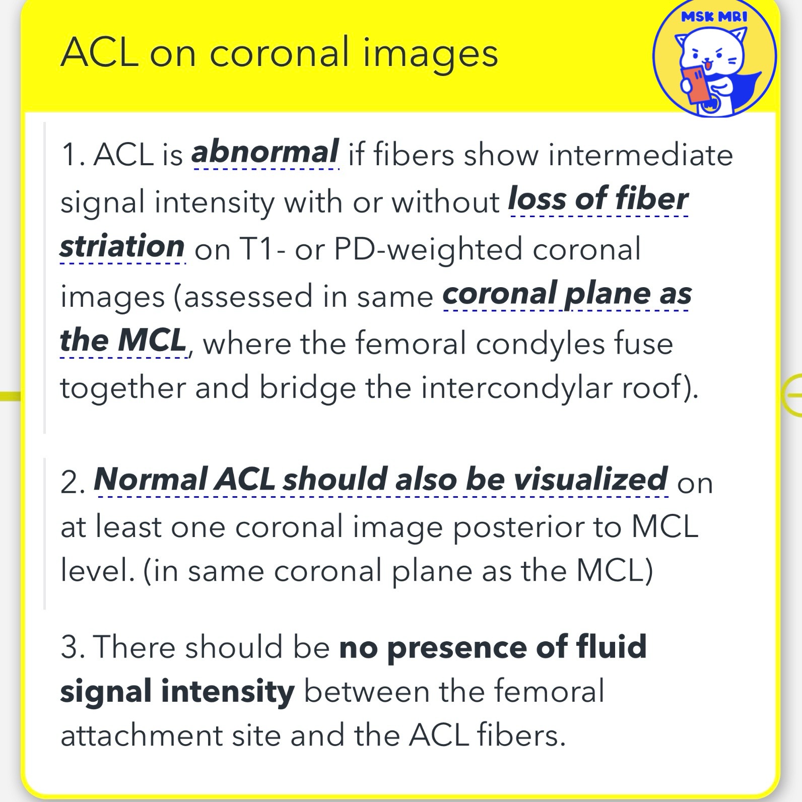
- In cases of an ACL tear at its femoral attachment, high signal intensity within the ACL and the presence of fluid between the torn ACL and the lateral femoral condyle are identifiable on coronal images.
- It's important to note that the normal ACL appears hypointense and maintains its ligamentous continuity on coronal images.
- This is specifically observed on at least one image posterior to, and including the plane of, the medial collateral ligament (MCL).
"Visualizing MSK Radiology: A Practical Guide to Radiology Mastery"
© 2022 MSK MRI Jee Eun Lee All Rights Reserved.
#VisualizingMSK #ACL #acltear #kneeMRI #emptynotchsign
You may not distribute or commercially exploit the content.
Nor may you transmit it or store it on any other website or other forms of the electronic retrieval system.
'✅ Knee MRI Mastery > Chap 2.ACL and PCL' 카테고리의 다른 글
| (Fig 2-B.07) Proximal ACL tear (0) | 2024.02.20 |
|---|---|
| (Fig 2-B.06) fluid between torn ACL and lateral femoral condyle sidewall -2_axial (0) | 2024.02.20 |
| (Fig 2-B.04) Empty notch sign (0) | 2024.02.19 |
| (Fig 2-B.02) Risk factors for ACL injuries increased slope of the tibial plateau. (0) | 2024.02.17 |
| (Fig 2-B.01) Risk factors for ACL injuries notch width index (1) | 2024.02.17 |


