==============================================
⬇️✨⬇️🎉⬇️🔥⬇️📚⬇️
Click the link to purchase on Amazon 🎉📚
==============================================
🎥 Check Out All Videos at Once! 📺
👉 Visit Visualizing MSK Blog to explore a wide range of videos! 🩻
https://visualizingmsk.blogspot.com/?view=magazine
📚 You can also find them on MSK MRI Blog and Naver Blog! 📖
https://www.instagram.com/msk_mri/
Click now to stay updated with the latest content! 🔍✨
==============================================
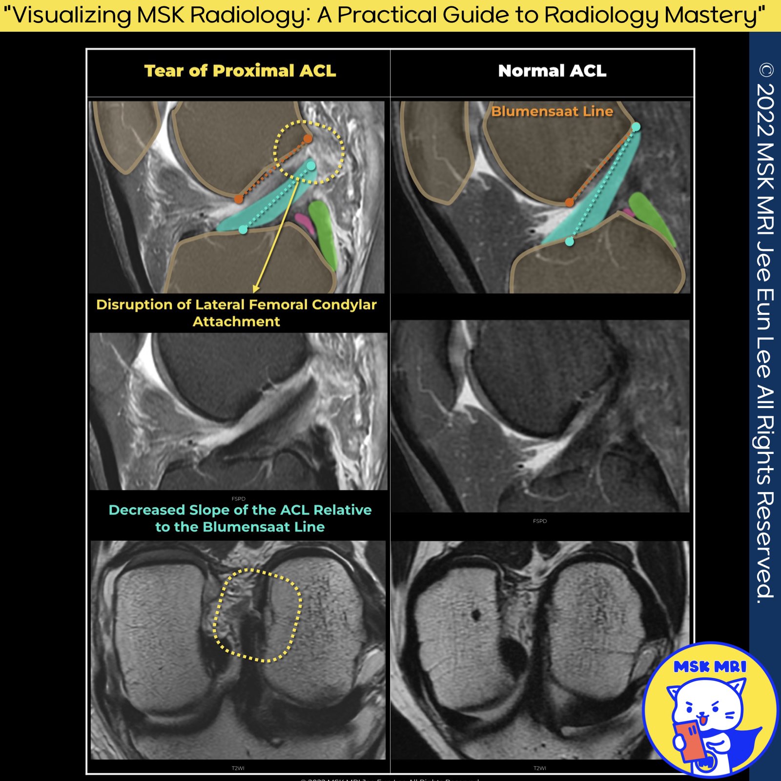
✅ Normal ACL vs. ACL Tear at Femoral Insertion
- Normal ACL:
Appears as an elliptical, homogeneous low signal on axial MRI images, indicating a firm attachment to the lateral femoral condyle. - ACL Tear at Femoral Insertion:
- Axial MRI: Shows increased signal within the proximal ACL fibers or fluid between the torn ACL and lateral femoral condyle, suggesting a tear.
- T2-Weighted Imaging: High signal intensity within the ACL and fluid presence are key indicators of a femoral attachment tear coronal images.
"Visualizing MSK Radiology: A Practical Guide to Radiology Mastery"
© 2022 MSK MRI Jee Eun Lee All Rights Reserved.
#VisualizingMSK #ACLinjuries #KneeMRI #ACLtear
You may not distribute or commercially exploit the content.
Nor may you transmit it or store it on any other website or other forms of the electronic retrieval system.
'✅ Knee MRI Mastery > Chap 2.ACL and PCL' 카테고리의 다른 글
| (Fig 2-B.09) Mass-like tissue without normally oriented ACL fibers (0) | 2024.02.20 |
|---|---|
| (Fig 2-B.08) Abnormal orientation or bowing of the ACL (0) | 2024.02.20 |
| (Fig 2-B.06) fluid between torn ACL and lateral femoral condyle sidewall -2_axial (0) | 2024.02.20 |
| (Fig 2-B.05) fluid between torn ACL and lateral femoral condyle sidewall -1_coronal (0) | 2024.02.20 |
| (Fig 2-B.04) Empty notch sign (0) | 2024.02.19 |


