==============================================
⬇️✨⬇️🎉⬇️🔥⬇️📚⬇️
Click the link to purchase on Amazon 🎉📚
==============================================
🎥 Check Out All Videos at Once! 📺
👉 Visit Visualizing MSK Blog to explore a wide range of videos! 🩻
https://visualizingmsk.blogspot.com/?view=magazine
📚 You can also find them on MSK MRI Blog and Naver Blog! 📖
https://www.instagram.com/msk_mri/
Click now to stay updated with the latest content! 🔍✨
==============================================
1️⃣ Importance in Tibial Tunnel Placement
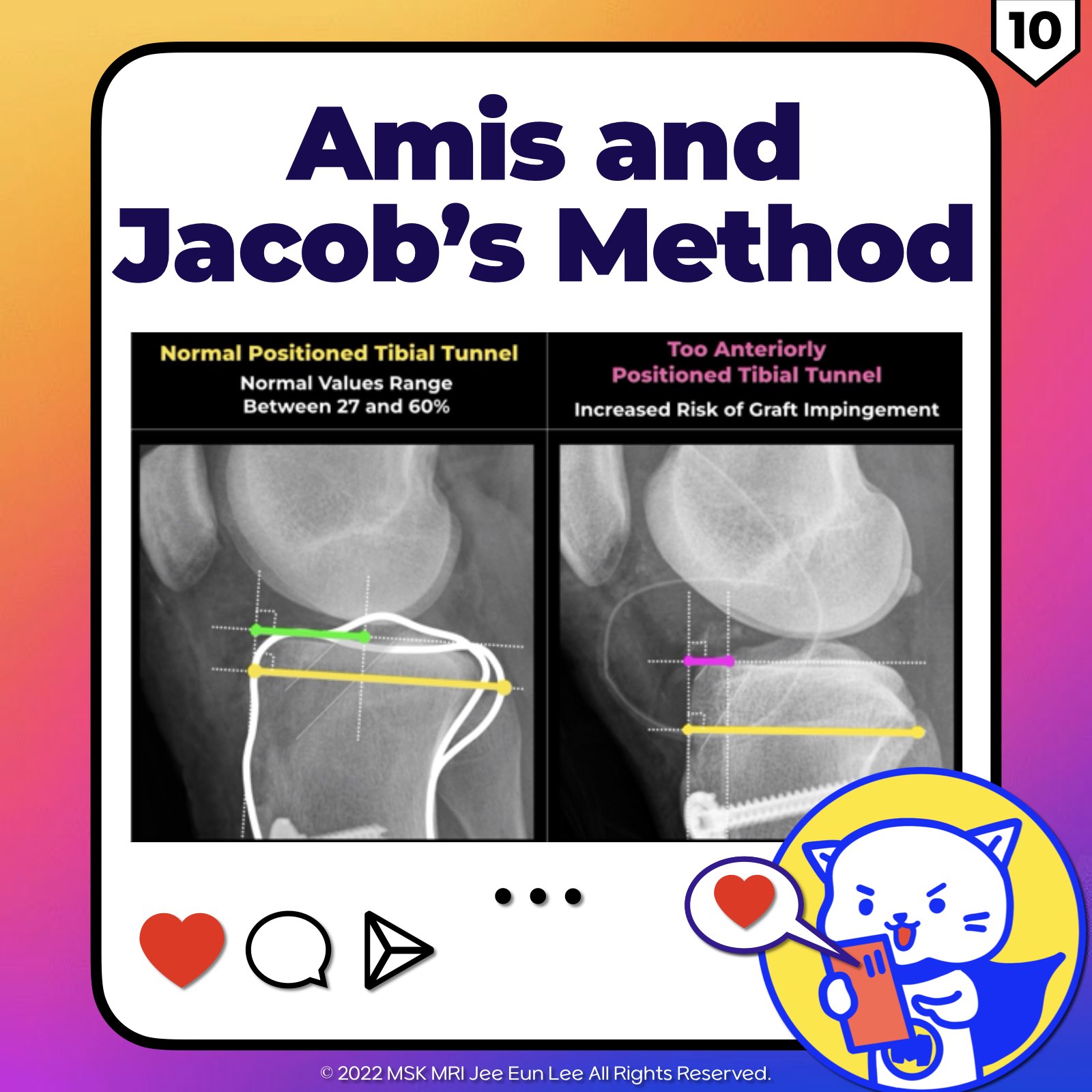
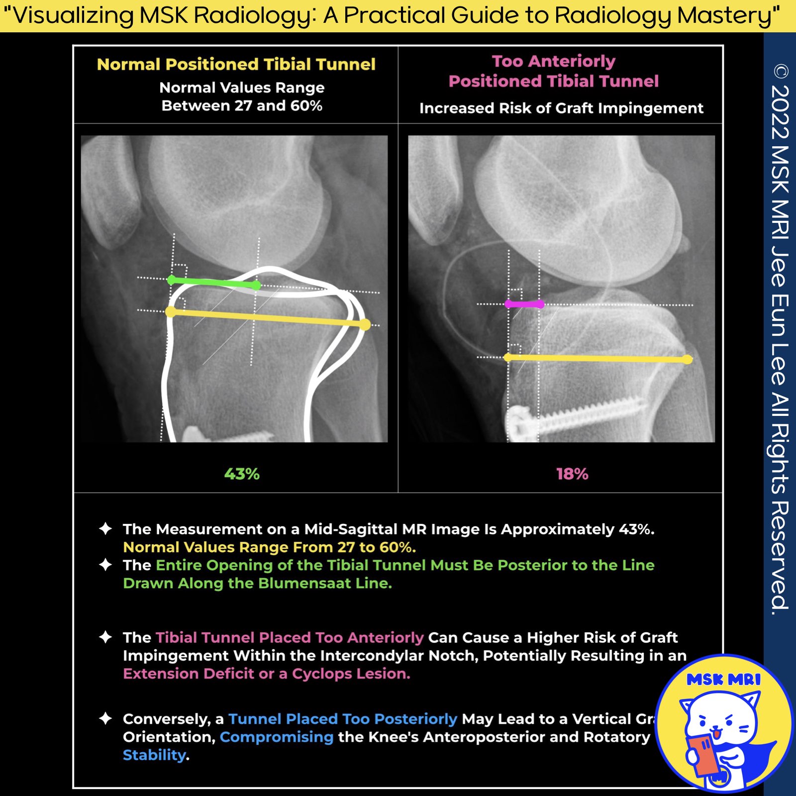
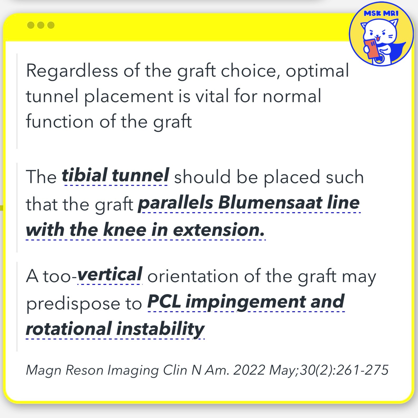
- The entire opening of the tibial tunnel must be located dorsally to the line drawn along the Blumensaat’s line.
2️⃣ Amis and Jakob's Line
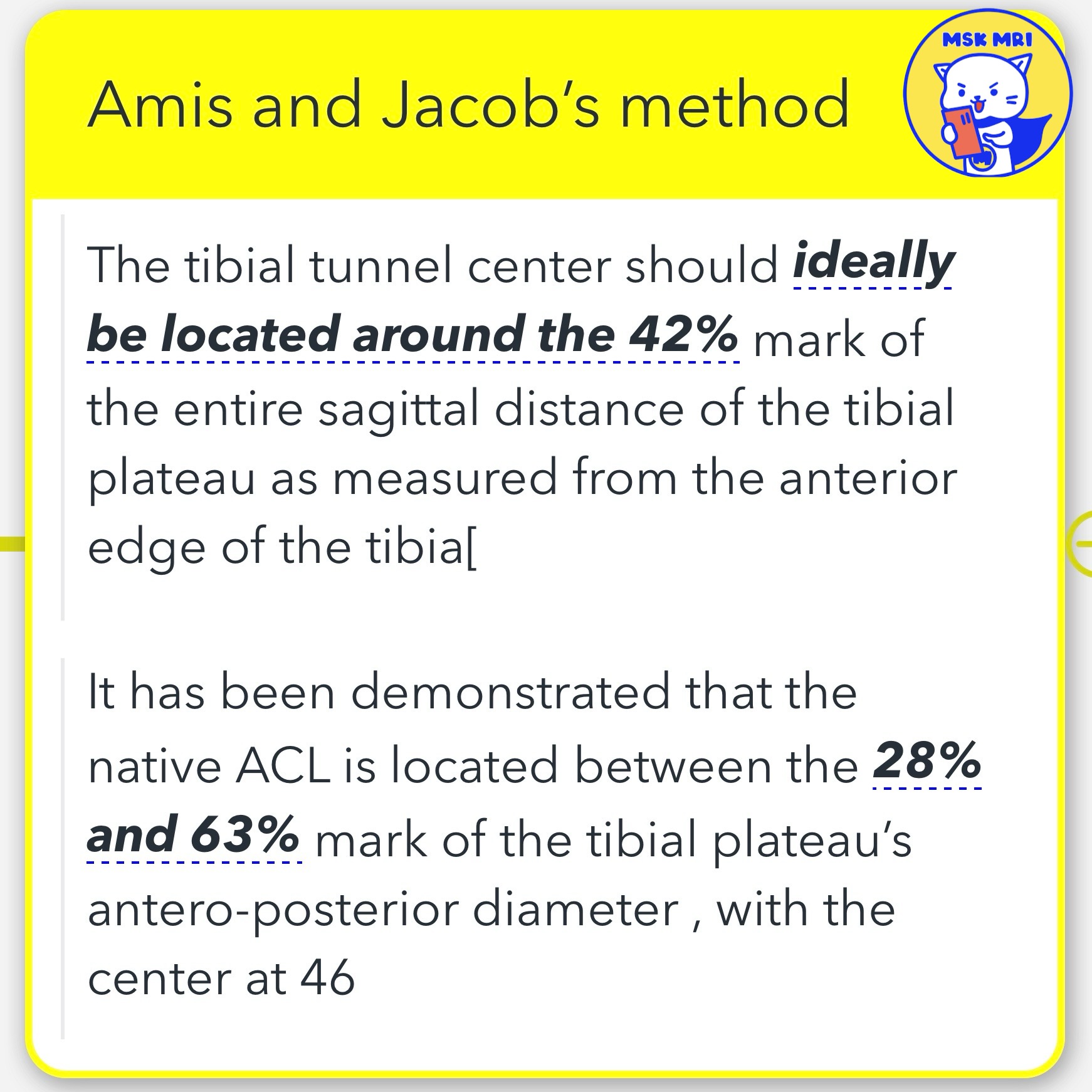
- The Amis and Jakob line is one of the most commonly used methods to evaluate the anterior-posterior direction of the tibial tunnel.
- The Amis and Jakob’s line is defined as a line passing through the most posterior edge of the tibial plateau and parallel to the medial tibial plateau surface.
- This method involves a percentage-term measurement generated from the Amis and Jakob’s line intersecting the anterior border (0%) and posterior border (100%) of the tibia plateau.
- The measurement is reported at around 43%. (Normal values range between 27 and 60%.)
3️⃣ Risks Associated with Incorrect Tunnel Placement
- If the tunnel is too forward, there is an increased risk of impingement of the graft with the intercondylar notch, possibly causing an extension deficit or a Cyclops lesion.
- Conversely, a tunnel positioned too posterior could lead to a vertical graft, responsible for an incomplete control of knee antero-posterior and rotatory stability.
"Visualizing MSK Radiology: A Practical Guide to Radiology Mastery" © 2022 MSK MRI Jee Eun Lee All Rights Reserved. #VisualizingMSK #ACLinjuries #KneeMRI #ACLtear #ACLreconstruction #tibialtunnel You may not distribute or commercially exploit the content. Nor may you transmit it or store it on any other website or other forms of the electronic retrieval system.
'✅ Knee MRI Mastery > Chap 2.ACL and PCL' 카테고리의 다른 글
| (Fig 2-C.13) MRI Characteristics of Different Graft Type (0) | 2024.03.03 |
|---|---|
| (Fig 2-C.12) ACL Graft Ligamentization (0) | 2024.03.03 |
| (Fig 2-C.09) Graft coronal Inclination angle (0) | 2024.03.02 |
| (Fig 2-C.07) Bi-dimensional sagittal inclination (0) | 2024.03.02 |
| (Fig 2-C.06) Bernard and Hertel’s grid-based technique (0) | 2024.03.02 |
