==============================================
⬇️✨⬇️🎉⬇️🔥⬇️📚⬇️
Click the link to purchase on Amazon 🎉📚
==============================================
🎥 Check Out All Videos at Once! 📺
👉 Visit Visualizing MSK Blog to explore a wide range of videos! 🩻
https://visualizingmsk.blogspot.com/?view=magazine
📚 You can also find them on MSK MRI Blog and Naver Blog! 📖
https://www.instagram.com/msk_mri/
Click now to stay updated with the latest content! 🔍✨
==============================================
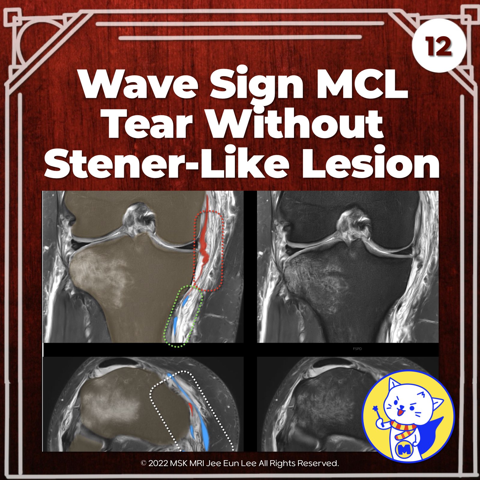
📌 The Wave Sign: Tears of the Distal Superficial Medial Collateral Ligament
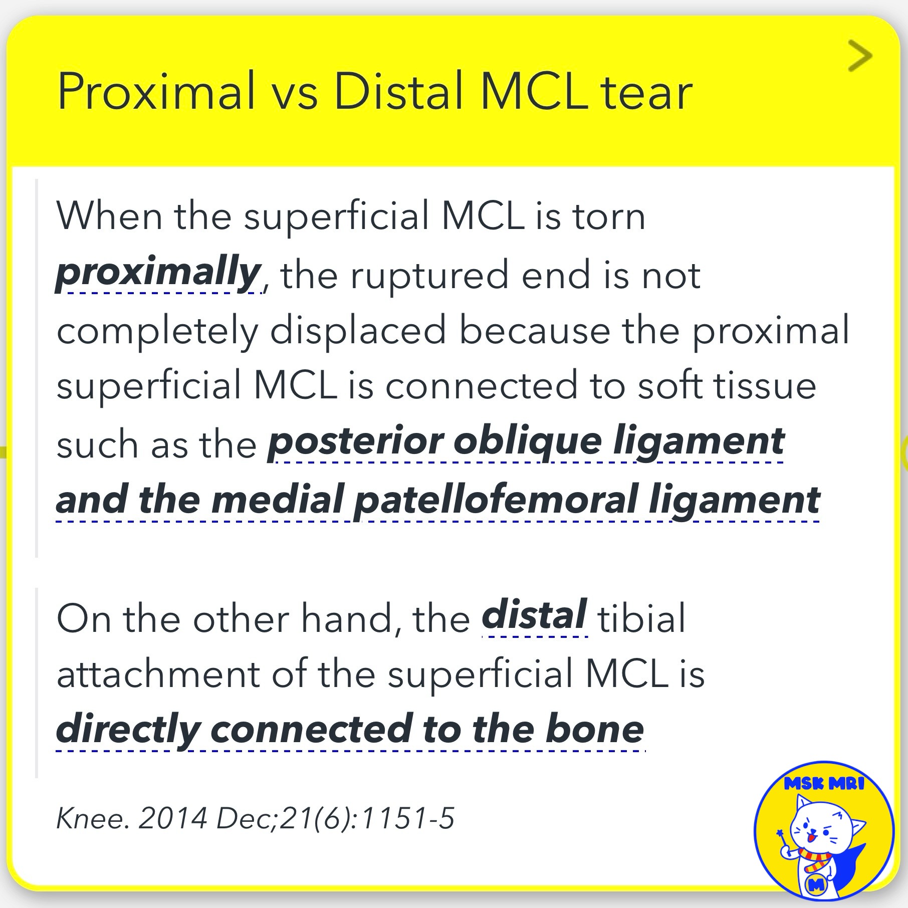
- Tears of the superficial medial collateral ligament (sMCL) most commonly involve the proximal or middle portion.
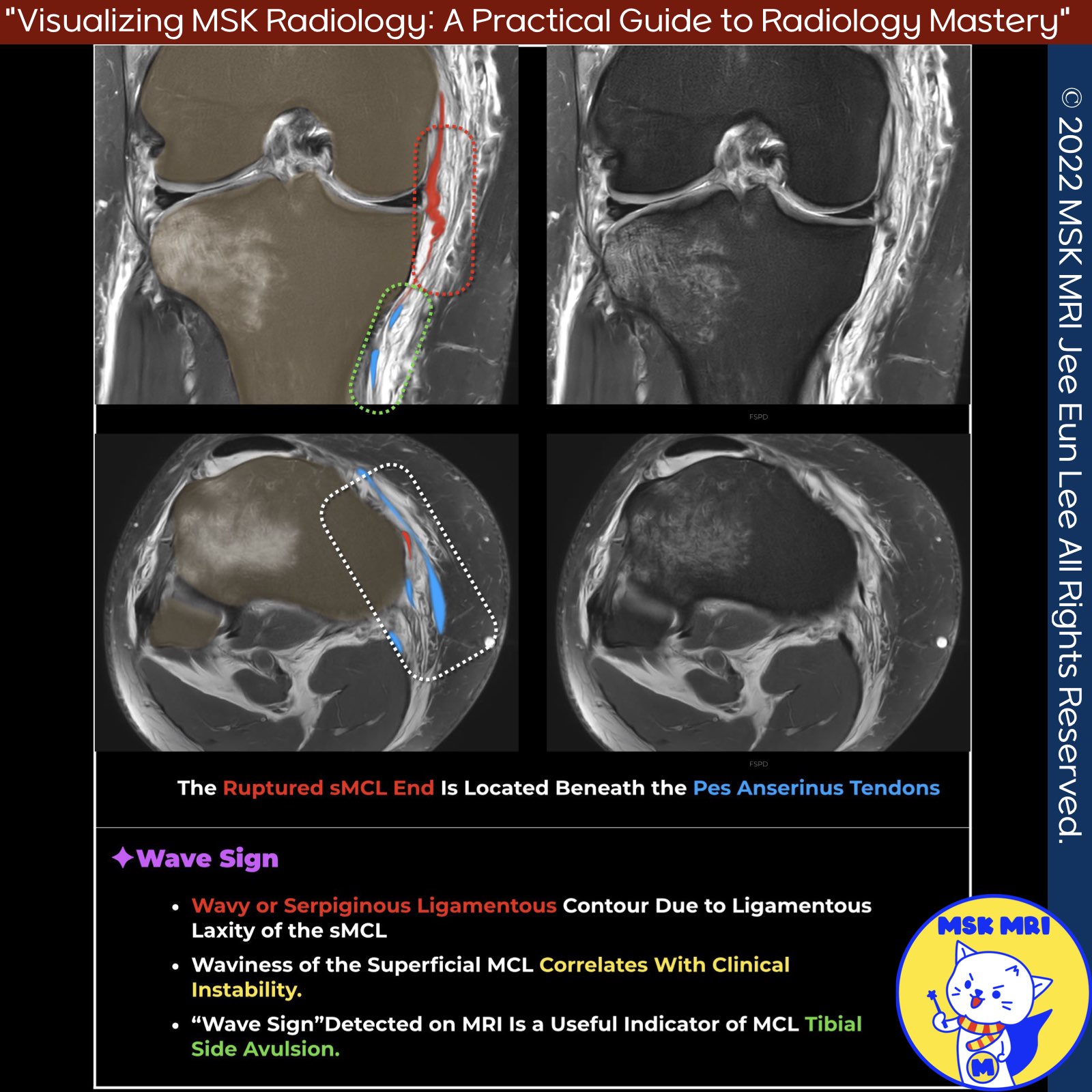
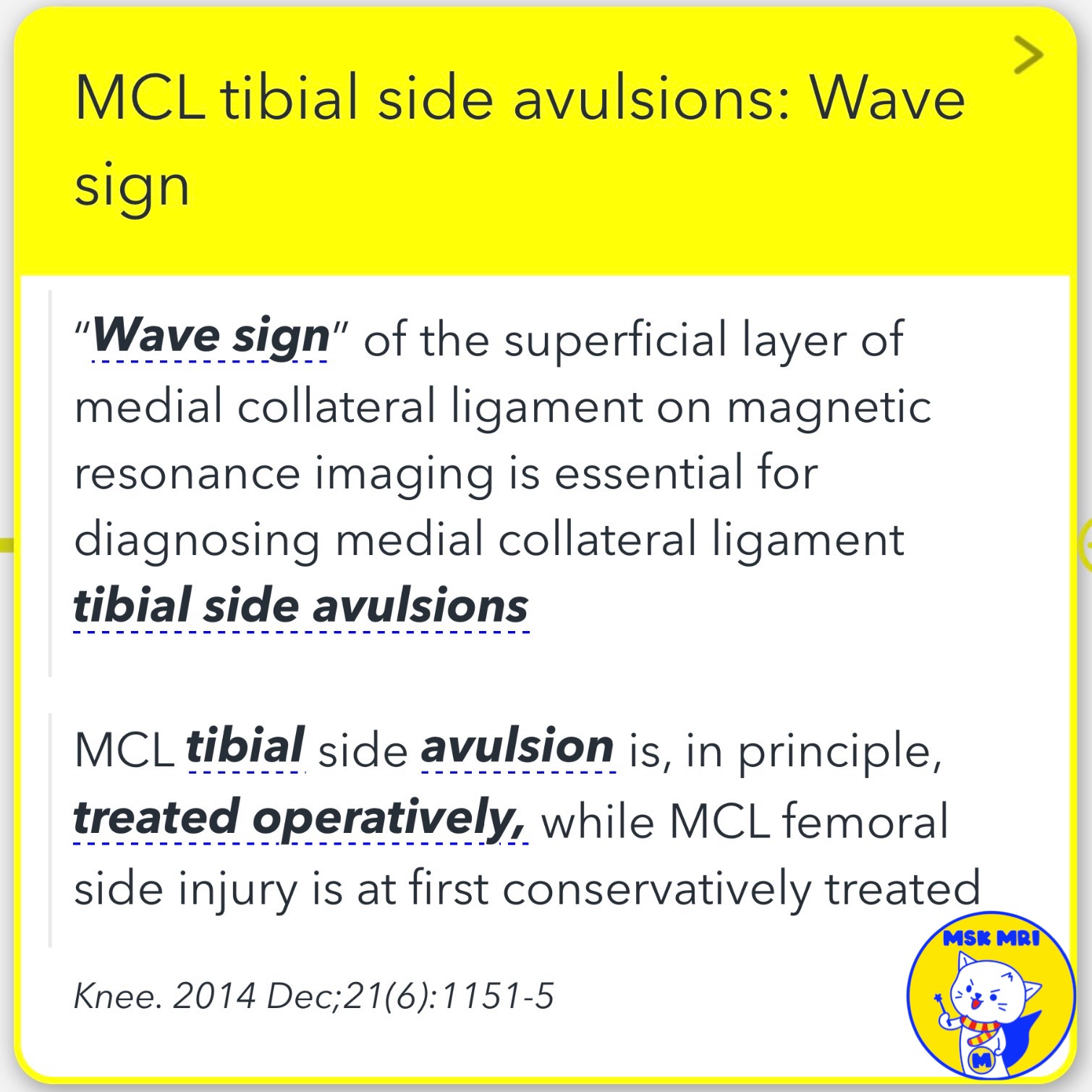
- The "wave sign" on MRI is essential for diagnosing medial collateral ligament tibial side avulsions.
- Waviness of the superficial MCL and injuries to other structures like the deep MCL, ACL, and posteromedial corner correlate with clinical instability.
✅ Differentiating from Pes Anserine Bursitis
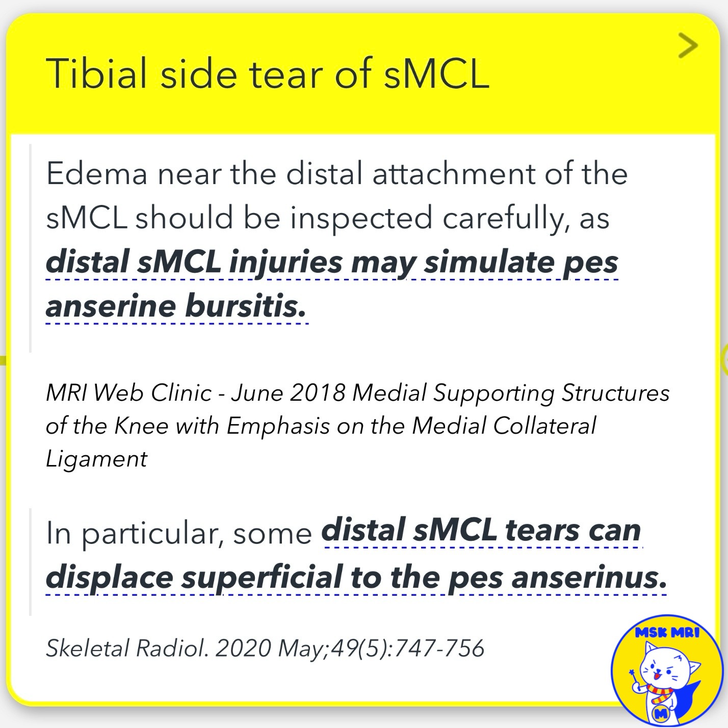
- Edema near the distal attachment of the sMCL should be carefully inspected, as distal sMCL injuries may simulate pes anserine bursitis.
- In pes anserine bursitis, the pes bursa is distended superficial to the MCL and deep to the pes anserinus tendons at the level of the proximal tibia, while the superficial MCL appears normal.
✅ Non-Stener-like Lesion
- Axial images should be examined carefully to determine if a Stener-like lesion has occurred.
- In a non-Stener-like lesion, the torn retracted ligament fibers of the distal superficial MCL remain deep to the pes anserinus tendons.
✅ Treatment Considerations
- MCL tibial side avulsion is typically treated operatively, while MCL femoral side injury is initially treated conservatively.
- Initial non-operative management is usually preferred for isolated grade III tears with an intact ACL.
Skeletal Radiol. 2020 May;49(5):747-756
Knee. 2014 Dec;21(6):1151-5
Semin Musculoskelet Radiol. 2016 Feb;20(1):12-25
"Visualizing MSK Radiology: A Practical Guide to Radiology Mastery"
© 2022 MSK MRI Jee Eun Lee All Rights Reserved.
No unauthorized reproduction, redistribution, or use for AI training.
#MCL, #sMCL, #MCLinjury, #Stenerlesion, #Pesanserine, #MCLtear,