==============================================
⬇️✨⬇️🎉⬇️🔥⬇️📚⬇️
Click the link to purchase on Amazon 🎉📚
==============================================
🎥 Check Out All Videos at Once! 📺
👉 Visit Visualizing MSK Blog to explore a wide range of videos! 🩻
https://visualizingmsk.blogspot.com/?view=magazine
📚 You can also find them on MSK MRI Blog and Naver Blog! 📖
https://www.instagram.com/msk_mri/
Click now to stay updated with the latest content! 🔍✨
==============================================
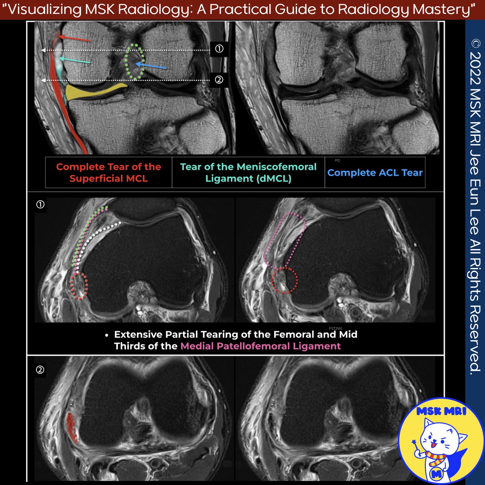
📌 MRI Findings in Grade 3 Superficial MCL Injury
- Complete discontinuity of the proximal superficial MCL and adjacent soft tissue edema, indicating a full-thickness tear.
📌 Involvement of MPFL in MCL Injuries
- Injuries of the superficial MCL can propagate anteriorly to involve the anterior MPFL.
- The MPFL merges with the fibers of the MCL at the epicondyle.
- The MPFL appears abnormal in most patients with clinically and MRI-documented cases of superficial MCL sprains or tears at or above the knee joint line.
✅ Clinical Relevance of MPFL Abnormalities
- The femoral third of the oblique decussation component of the MPFL, which arises from the anterior margin of the upper superficial MCL, appears abnormal in approximately 90% of cases involving superficial MCL injuries.
- Despite these MPFL abnormalities, there is no evidence of concurrent lateral patellar dislocation on the initial MRI scans.
✅ MPFL Anatomy
- The MPFL consists of a thinner, more superior transverse component attaching to the medial femoral condyle and a broader, more inferior oblique decussation component arising from the upper segment of the superficial MCL.
Skeletal Radiol. 2022 Jul;51(7):1381-1389.
"Visualizing MSK Radiology: A Practical Guide to Radiology Mastery"
© 2022 MSK MRI Jee Eun Lee All Rights Reserved.
No unauthorized reproduction, redistribution, or use for AI training.
#MCL, #sMCL, #MCLinjury, #Valgusinjury, #MCLtear, #MPFL, #MPFLinjury,