Click the link to purchase on Amazon 🎉📚
==============================================
🎥 Check Out All Videos at Once! 📺
👉 Visit Visualizing MSK Blog to explore a wide range of videos! 🩻
https://visualizingmsk.blogspot.com/?view=magazine
📚 You can also find them on MSK MRI Blog and Naver Blog! 📖
https://www.instagram.com/msk_mri/
Click now to stay updated with the latest content! 🔍✨
==============================================
📌Characteristics of Distal MCL Tears
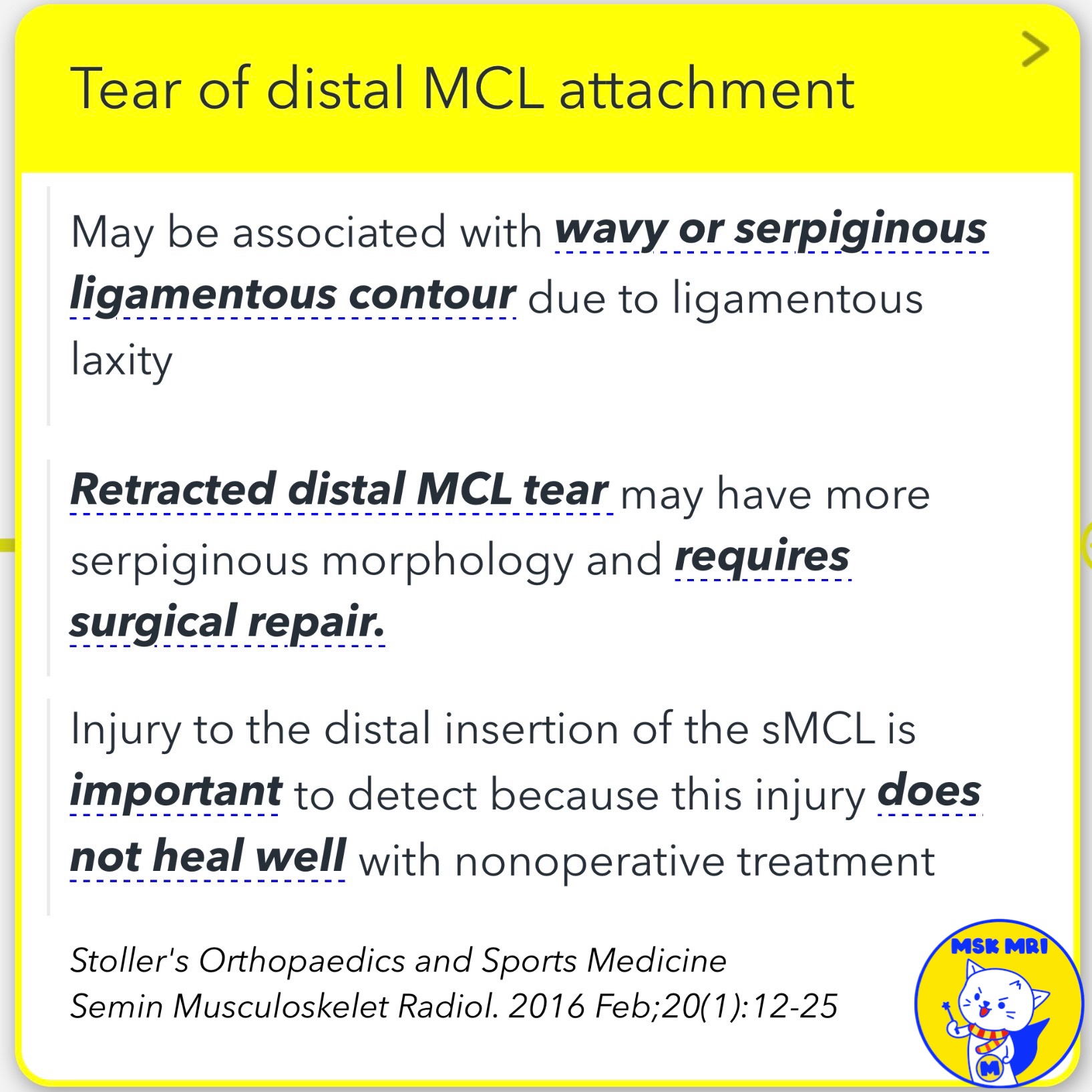
- May have a wavy or serpiginous ligamentous contour due to laxity
- Retracted tears may have a more serpiginous morphology, requiring surgical repair
- Distal sMCL insertion injuries do not heal well with nonoperative treatment
📌Proximal vs. Distal MCL Tear Differences
- In proximal sMCL tears, the ruptured end is not completely displaced due to connections to surrounding soft tissues
- Distal sMCL tears have a direct bone attachment, with impaired healing due to poor blood supply, lack of soft tissue connections, and potential displacement
✅ Management Approach
- Distal MCL tibial side avulsions are treated operatively
- Proximal MCL femoral side injuries are initially treated conservatively
➡️ [Case]
- Distal sMCL attachment is broad, 6-7 cm below the joint line and deep to the pes anserine tendons
- Case shows distal sMCL tear with laxity and MCL bursitis
- Pes anserine tendons (sartorius, gracilis, semitendinosus) attach anteromedially on the tibia
- Axial images are crucial to determine if the sMCL stump is deep (non-displaced) or superficial (displaced) to the pes anserinus
- This case represents a non-Stener-like lesion, with the torn sMCL fibers remaining deep to the pes anserinus
Stoller's Orthopaedics and Sports Medicine
Semin Musculoskelet Radiol. 2016 Feb;20(1):12-25
Knee. 2014 Dec;21(6):1151-5
Skeletal Radiol. 2020 May;49(5):747-756
"Visualizing MSK Radiology: A Practical Guide to Radiology Mastery"
© 2022 MSK MRI Jee Eun Lee All Rights Reserved.
No unauthorized reproduction, redistribution, or use for AI training.
#MCL, #sMCL, #MCLinjury, #Stenerlesion, #Pesanserine, #MCLtear,


