Click the link to purchase on Amazon 🎉📚
==============================================
🎥 Check Out All Videos at Once! 📺
👉 Visit Visualizing MSK Blog to explore a wide range of videos! 🩻
https://visualizingmsk.blogspot.com/?view=magazine
📚 You can also find them on MSK MRI Blog and Naver Blog! 📖
https://www.instagram.com/msk_mri/
Click now to stay updated with the latest content! 🔍✨
==============================================
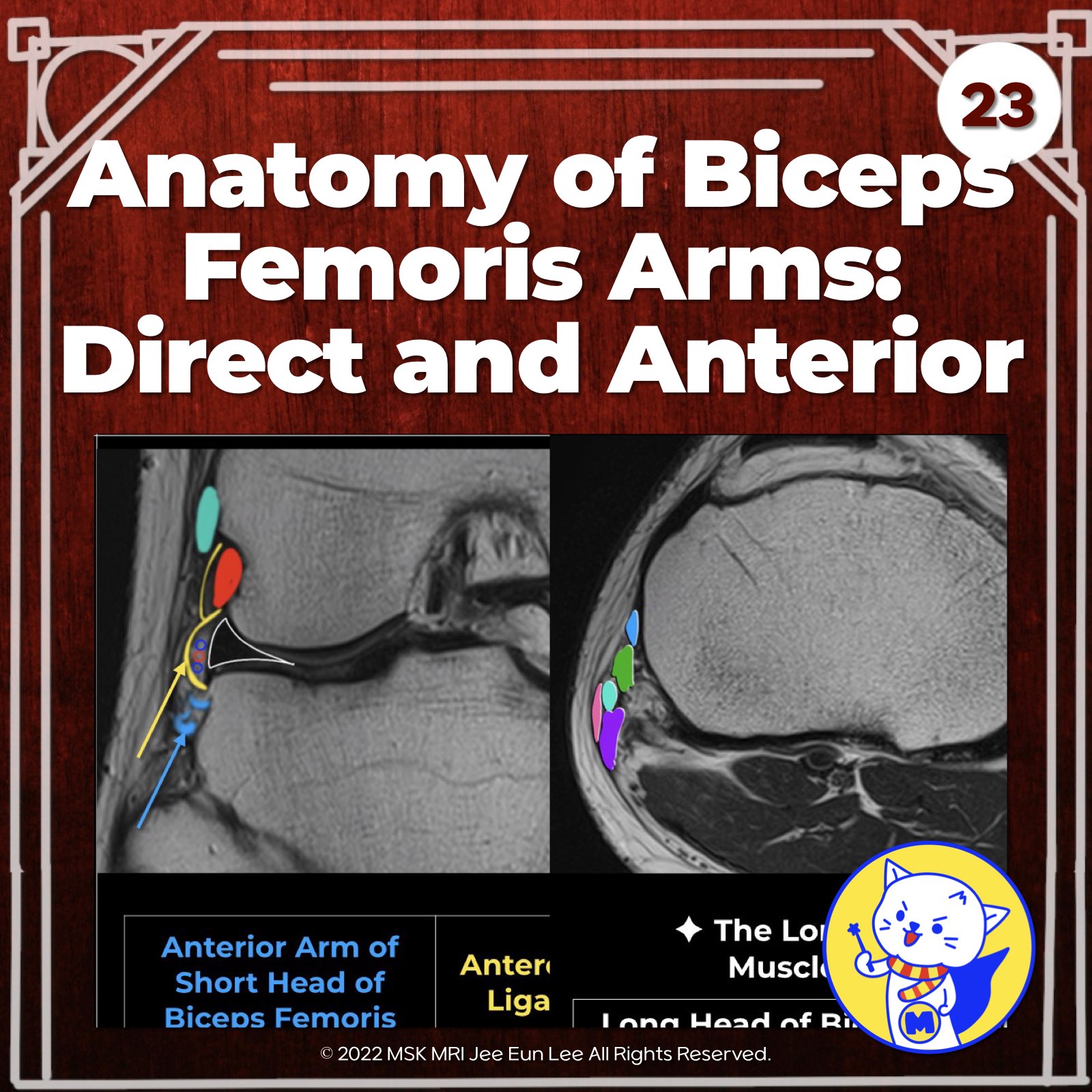
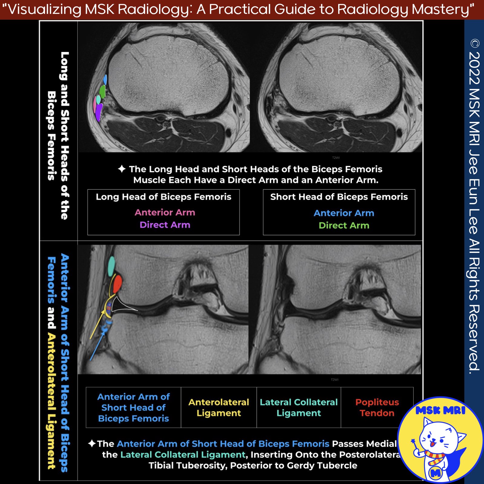
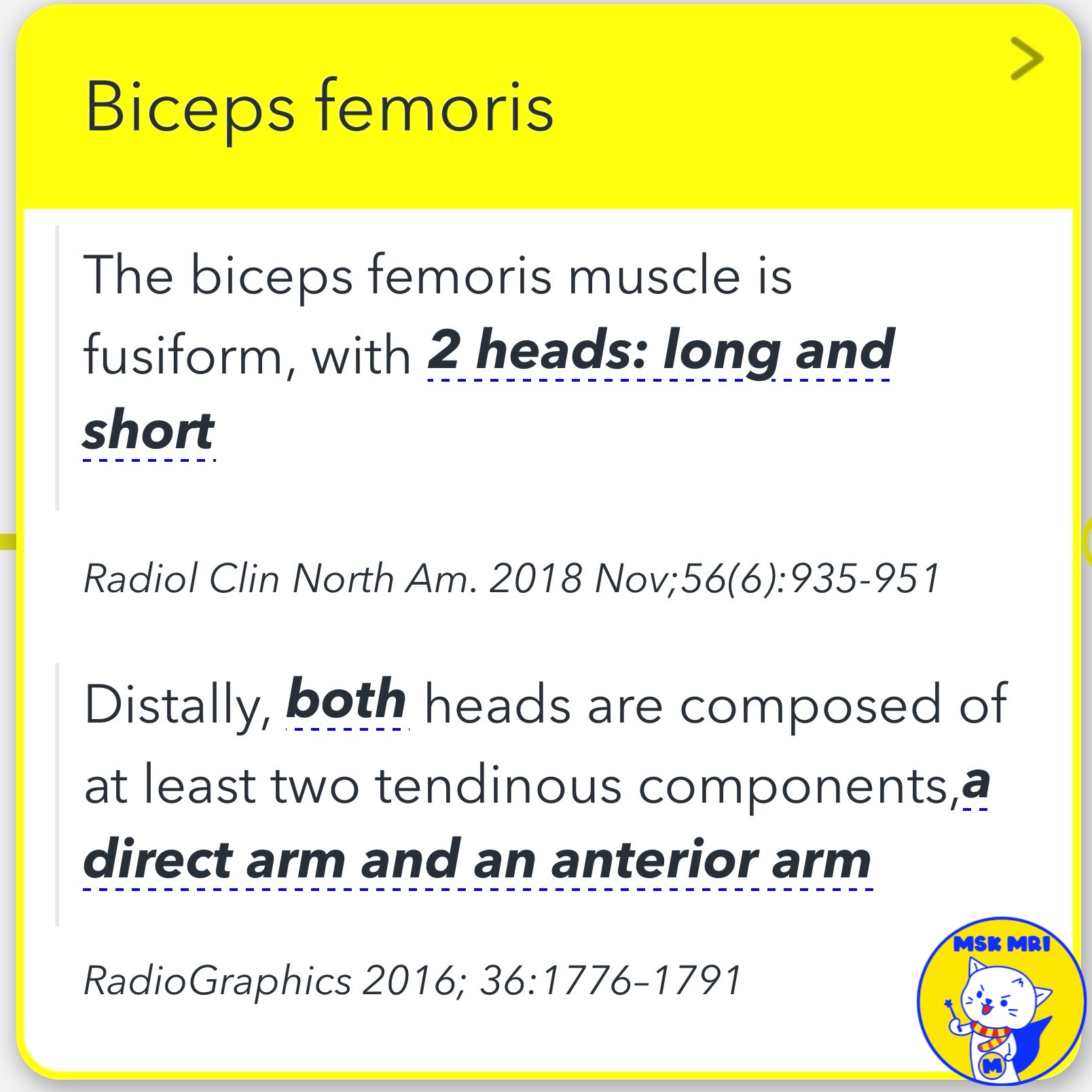
📌 Anatomy of the Biceps Femoris Tendon
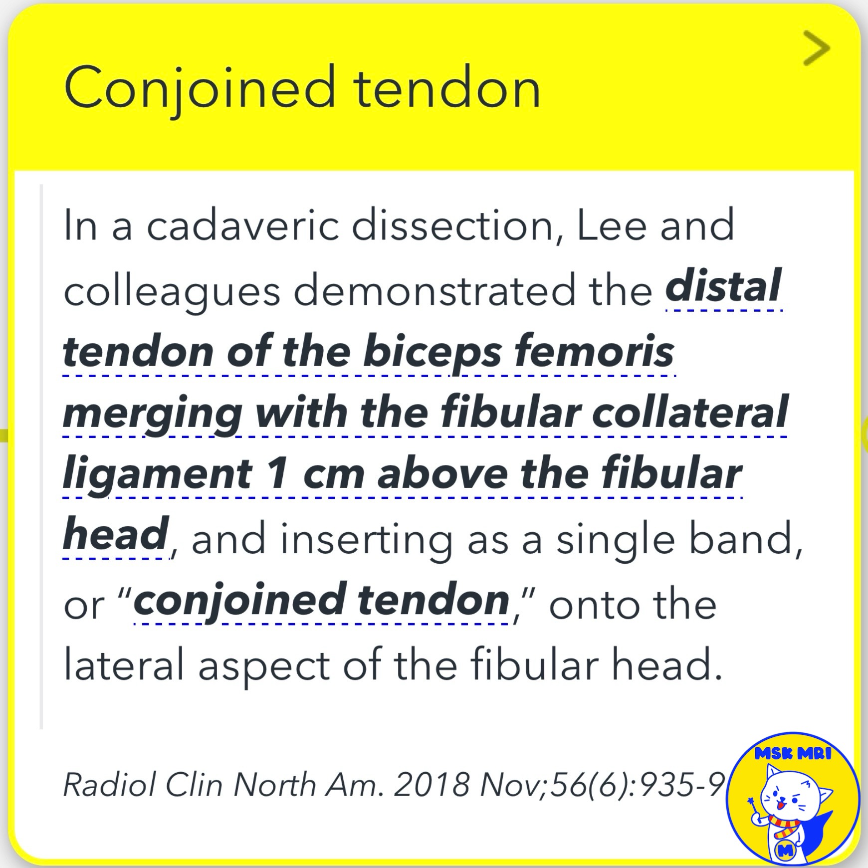
- Distal Biceps Femoris Tendon The distal tendon of the biceps femoris merges with the lateral collateral ligament (LCL) 1 cm above the fibular head, inserting as a single conjoined tendon onto the lateral aspect of the fibular head.
- The LCL inserts anteriorly and the biceps femoris tendon inserts posteriorly.
- Tendon Composition on Axial Imaging On axial imaging, the biceps femoris tendon does not appear as a single structure.
- Instead, it consists of different tendinous and fascial components, most attaching to the fibular head, with potential tibial attachment.
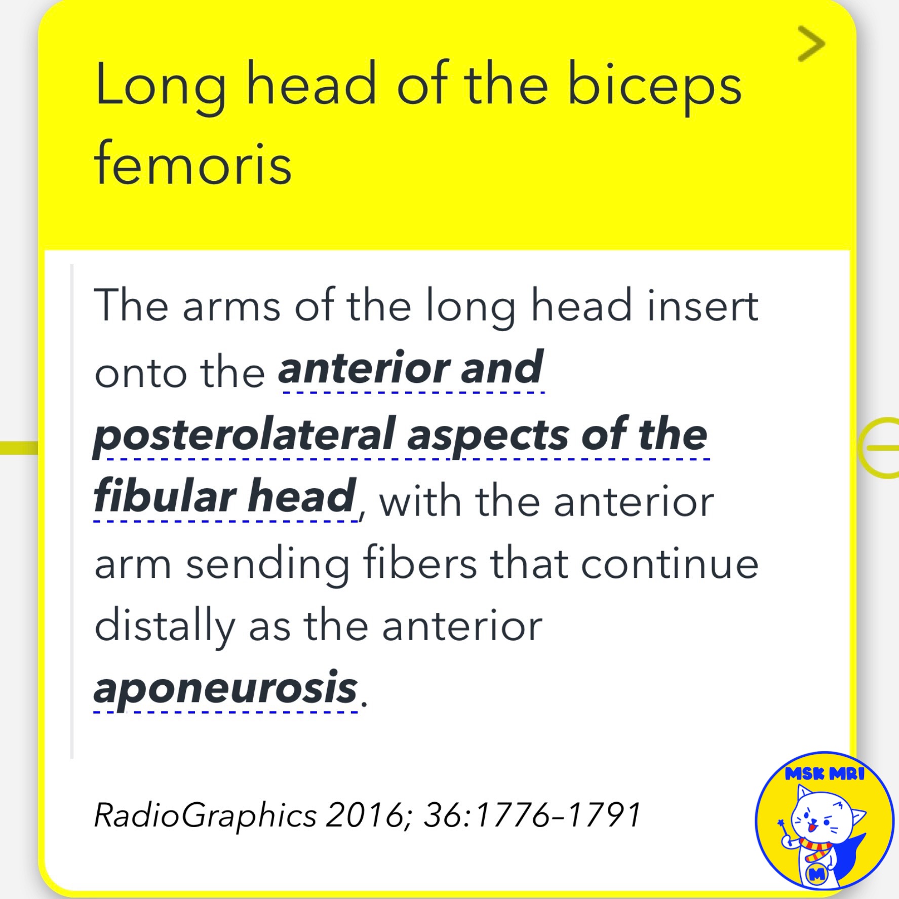
✅Long Head Tendon Arms The long head
- Anterior Arm - Inserts on lateral fibular head/neck
- Direct Arm - Inserts posteriorly on lateral fibular head/neck
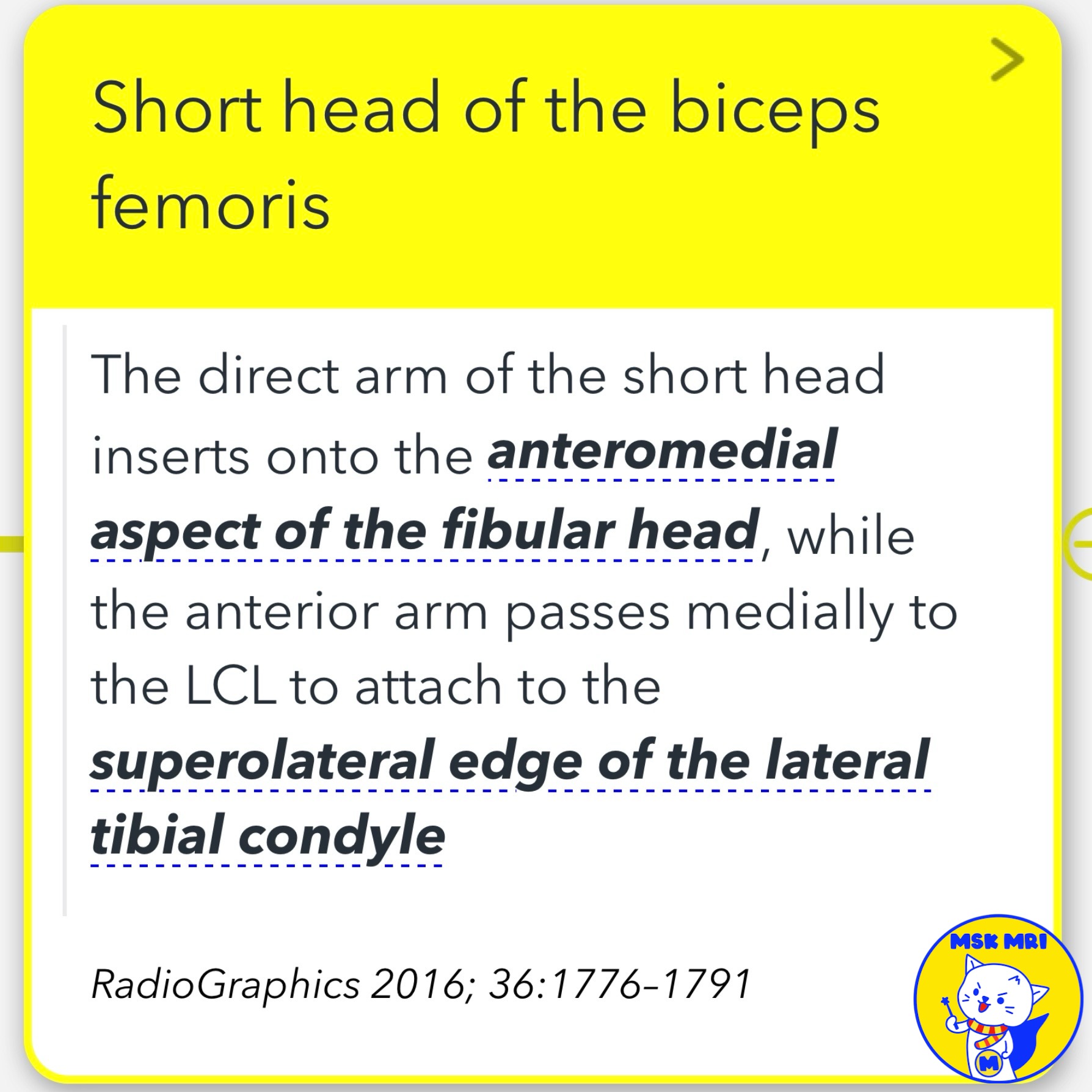
✅ Short Head Tendon Arms
- Direct Arm - Inserts anteromedially on fibular head
- Anterior Arm - Does not attach to fibula, passes medial to LCL to attach on superolateral tibial condyle
★ Additional Point
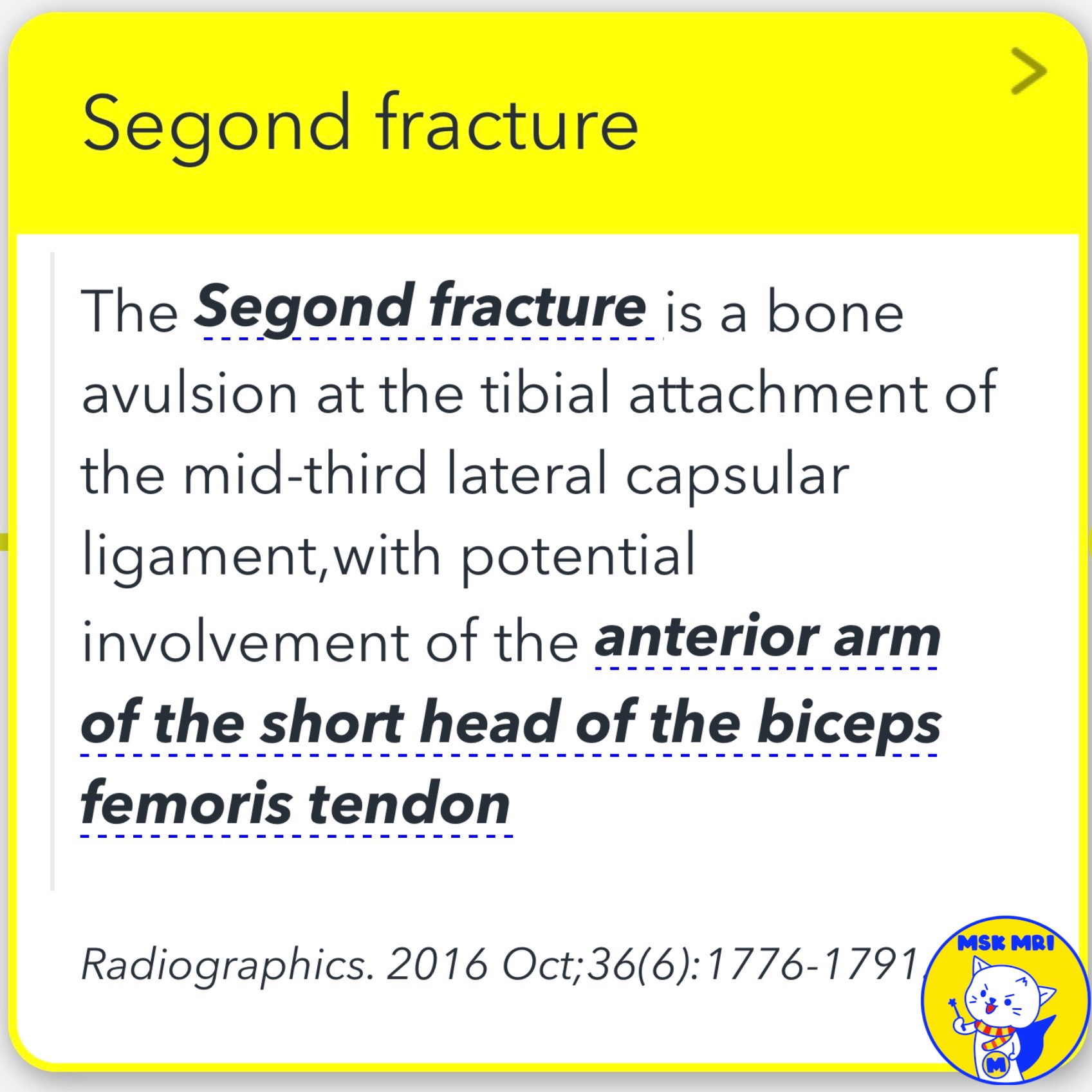
- The anterior arm of the short head attaches posterior to Gerdy's tubercle, distal to the anterolateral ligament insertion.
- Therefore, a Segond fracture (avulsion of the mid-third lateral capsular ligament) may involve this tendon arm.
Radiol Clin North Am. 2018 Nov;56(6):935-951
RadioGraphics 2016; 36:1776–1791
Radiographics. 2016 Oct;36(6):1776-1791.
"Visualizing MSK Radiology: A Practical Guide to Radiology Mastery"
© 2022 MSK MRI Jee Eun Lee All Rights Reserved.
No unauthorized reproduction, redistribution, or use for AI training.
#bicepsfemoristendon, #bicepsfemorisanatomy, #kneetendonanatomy, #lateralcollateralligament, #LCLanatomy, #fibularheadanatomy, #tibialcondyleanatomy, #segondfracture, #anterolateralligament,
'✅ Knee MRI Mastery > Chap 3.Collateral Ligaments' 카테고리의 다른 글
| (Fig 3-B.25) Biceps Femoris Tendinosis and Associated Bursitis (0) | 2024.05.22 |
|---|---|
| (Fig 3-B.24) Distal Biceps Femoris Myotendinous Junction Tear (0) | 2024.05.22 |
| (Fig 3-B.22) Arcuate and Fabellofibular Ligament Tears (0) | 2024.05.22 |
| (Fig 3-B.20) Arcuate Ligament Anatomy (1) | 2024.05.22 |
| (Fig 3-B.19) Fabellofibular Ligament Anatomy (0) | 2024.05.22 |