Click the link to purchase on Amazon 🎉📚
==============================================
🎥 Check Out All Videos at Once! 📺
👉 Visit Visualizing MSK Blog to explore a wide range of videos! 🩻
https://visualizingmsk.blogspot.com/?view=magazine
📚 You can also find them on MSK MRI Blog and Naver Blog! 📖
https://www.instagram.com/msk_mri/
Click now to stay updated with the latest content! 🔍✨
==============================================
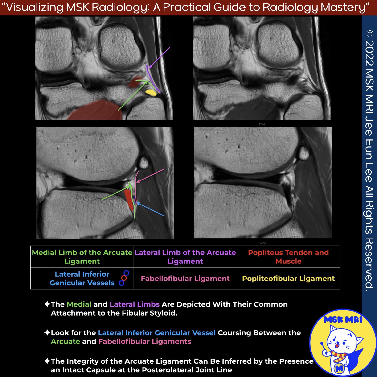
📌 Anatomy of the Arcuate Ligament
- Y-shaped thickening of the posterolateral capsule
- Runs to apex of fibular styloid process
- Lateral limb attaches to lateral femoral condyle and posterolateral joint capsule
- Medial limb attaches to posterior knee capsule and oblique popliteal ligament
✅ Variability and Visualization
- Variably present structure
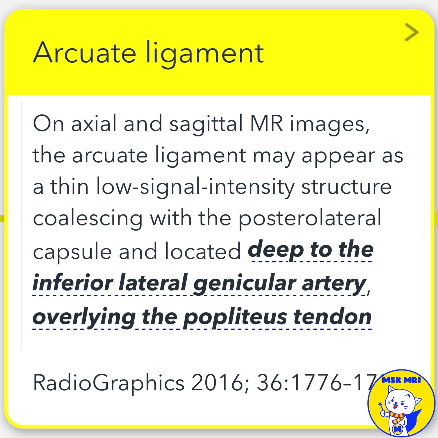
- Difficult to visualize directly on MRI even when present
✅ Key Point 1: No Need to Decipher Exact Anatomy
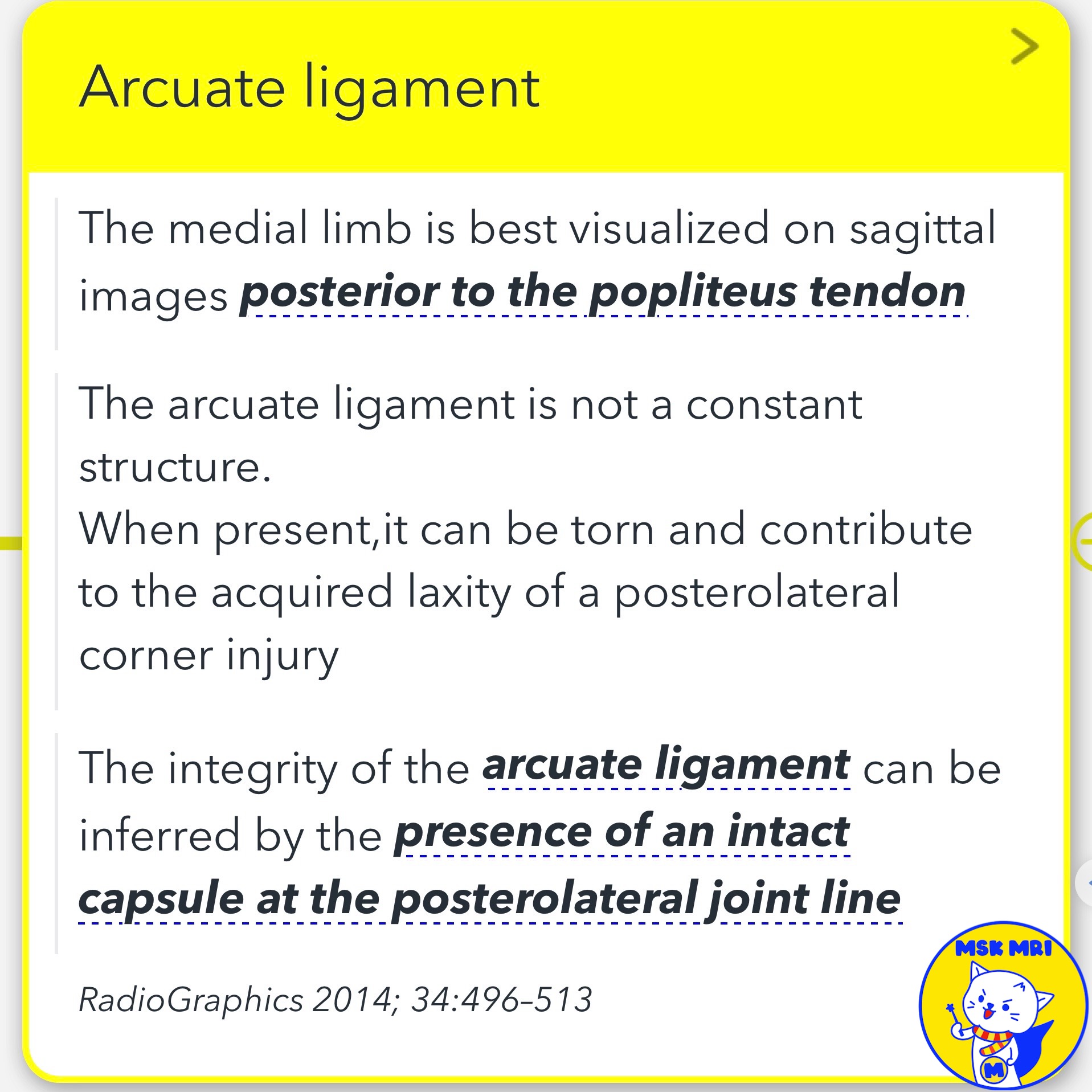
- Injury can be inferred from increased pericapsular signal around posterolateral capsule
- Torn medial limb seen posterior to popliteus tendon
- Integrity inferred from intact posterolateral joint line capsule
✅ Key Point 2: Sagittal Anatomy to Remember
- Popliteus tendon lies deepest, easy to identify
- Lateral inferior genicular vessel also easily seen
- Medial limb between popliteus and genicular vessel
- Fabellofibular ligament superficial to genicular vessel, connects to fabella
- If fabellofibular ligament prominent, lateral limb may be small/absent
RadioGraphics 2016; 36:1776–1791
RadioGraphics 2014; 34:496–513
Stoller's Orthopaedics and Sports Medicine: The Knee
"Visualizing MSK Radiology: A Practical Guide to Radiology Mastery"
© 2022 MSK MRI Jee Eun Lee All Rights Reserved.
No unauthorized reproduction, redistribution, or use for AI training.
#ArcuateLigamentAnatomy, #PosterolateralKneeCapsule, #YShapedLigament, #LateralLimb, #MedialLimb, #PopliteusTendon, #LateralInferiorGeniculaVessel, #FabellofibularLigament,
#Knee, #MRI, #Anatomy, #Ligaments, #PosterolateralCorner, #ArcuateLigament, #PopliteusTendon, #GeniculaVessels, #FabellofibularLigament, #CapsuleIntegrity


'✅ Knee MRI Mastery > Chap 3.Collateral Ligaments' 카테고리의 다른 글
| (Fig 3-B.23) Anatomy of Biceps Femoris Arms: Direct and Anterior (0) | 2024.05.22 |
|---|---|
| (Fig 3-B.22) Arcuate and Fabellofibular Ligament Tears (0) | 2024.05.22 |
| (Fig 3-B.19) Fabellofibular Ligament Anatomy (0) | 2024.05.22 |
| (Fig 3-B.18) Popliteofibular Ligament Avulsion Fracture (0) | 2024.05.22 |
| (Fig 3-B.17) Partial Popliteofibular Ligament Tear (0) | 2024.05.22 |