Click the link to purchase on Amazon 🎉📚
==============================================
🎥 Check Out All Videos at Once! 📺
👉 Visit Visualizing MSK Blog to explore a wide range of videos! 🩻
https://visualizingmsk.blogspot.com/?view=magazine
📚 You can also find them on MSK MRI Blog and Naver Blog! 📖
https://www.instagram.com/msk_mri/
Click now to stay updated with the latest content! 🔍✨
==============================================
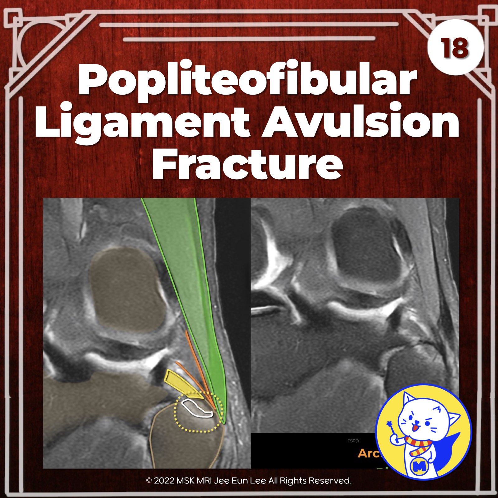
📌Attachments to the Fibular Head
- Biceps femoris tendon and lateral collateral ligament attach laterally
- Arcuate and fabellofibular ligaments attach posteromedially
- Popliteofibular ligament attaches most medially
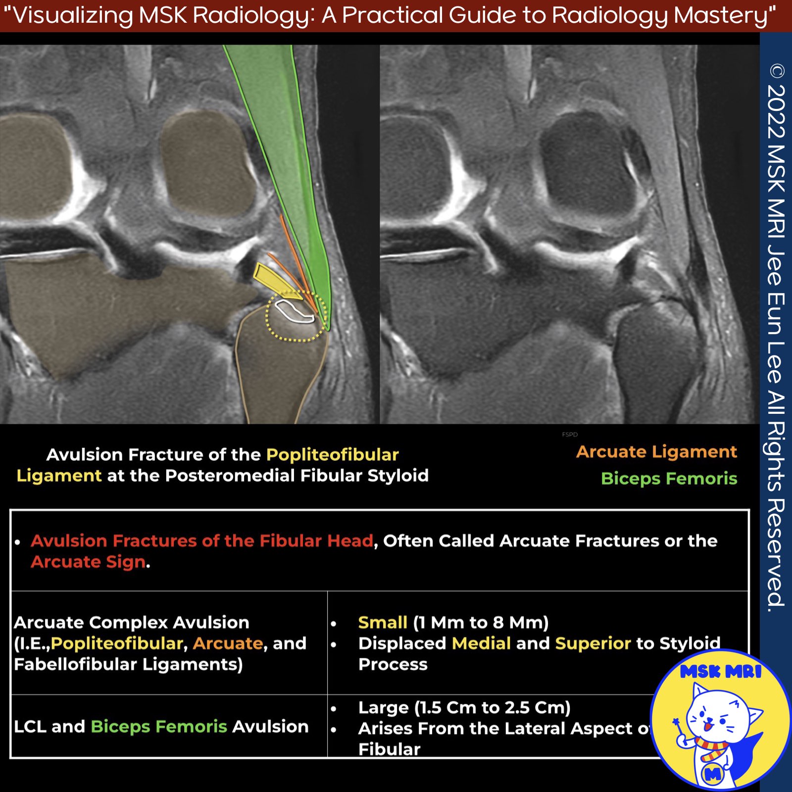
✅ Fibular Head Fractures as Clues to PLC Injury
- Avulsion fractures of the fibular head (arcuate fractures/sign) occur at PLC component attachment sites
- Their location and size help predict the injured structure
✅ Fracture Patterns and Injured Structures
- Diffuse/lateral fibular head fracture = fibular collateral ligament or biceps femoris injury
- Small styloid fracture = popliteofibular or arcuate ligament injury
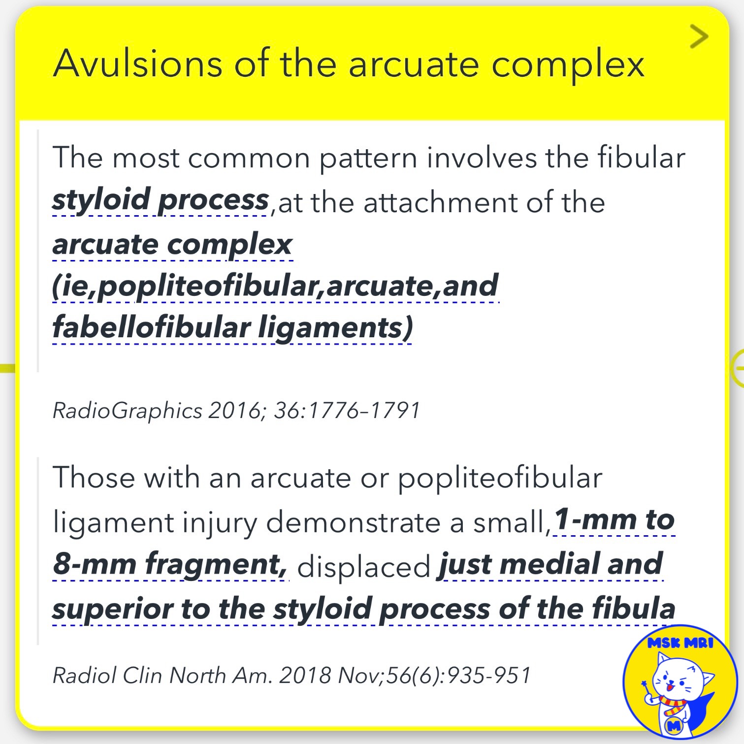
✅ Arcuate Complex Avulsions
- Common pattern involves styloid process at arcuate complex attachment
- 1-8mm fragment displaced medial/superior to styloid = arcuate or popliteofibular ligament injury
RadioGraphics 2016; 36:1776–1791
RadioGraphics 2014; 34:496–513
Radiol Clin North Am. 2018 Nov;56(6):935-951
"Visualizing MSK Radiology: A Practical Guide to Radiology Mastery"
© 2022 MSK MRI Jee Eun Lee All Rights Reserved.
No unauthorized reproduction, redistribution, or use for AI training.
#posterolateralcornerinjury, #fibularheadfracture, #arcuatefracture, #arcuatesign, #popliteofibularligamentinjury, #arcuateligamentinjury, #fabellofibularligamentinjury, #lateralcollateralligamentinjury, #bicepsfemoristendoninjury, #avulsionfracture

'✅ Knee MRI Mastery > Chap 3.Collateral Ligaments' 카테고리의 다른 글
| (Fig 3-B.20) Arcuate Ligament Anatomy (1) | 2024.05.22 |
|---|---|
| (Fig 3-B.19) Fabellofibular Ligament Anatomy (0) | 2024.05.22 |
| (Fig 3-B.17) Partial Popliteofibular Ligament Tear (0) | 2024.05.22 |
| (Fig 3-B.16) Popliteofibular Ligament Anatomy (0) | 2024.05.22 |
| (Fig 3-B.15) Popliteus Tendon Avulsion Fracture (0) | 2024.05.21 |