Click the link to purchase on Amazon 🎉📚
==============================================
🎥 Check Out All Videos at Once! 📺
👉 Visit Visualizing MSK Blog to explore a wide range of videos! 🩻
https://visualizingmsk.blogspot.com/?view=magazine
📚 You can also find them on MSK MRI Blog and Naver Blog! 📖
https://www.instagram.com/msk_mri/
Click now to stay updated with the latest content! 🔍✨
==============================================
📌 Popliteofibular Ligament
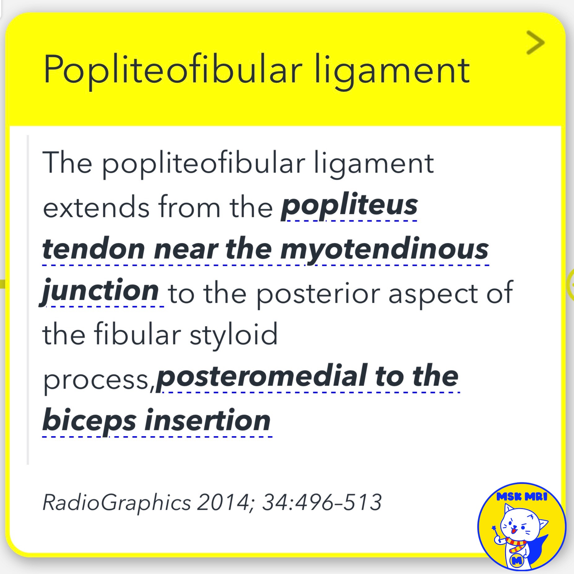
The popliteofibular ligament is a sturdy tendinous band that crucially stabilizes the posterolateral corner of the knee.
It extends from the popliteus tendon to the posterior aspect of the fibular styloid process.
★ Identifying the Popliteofibular Ligament
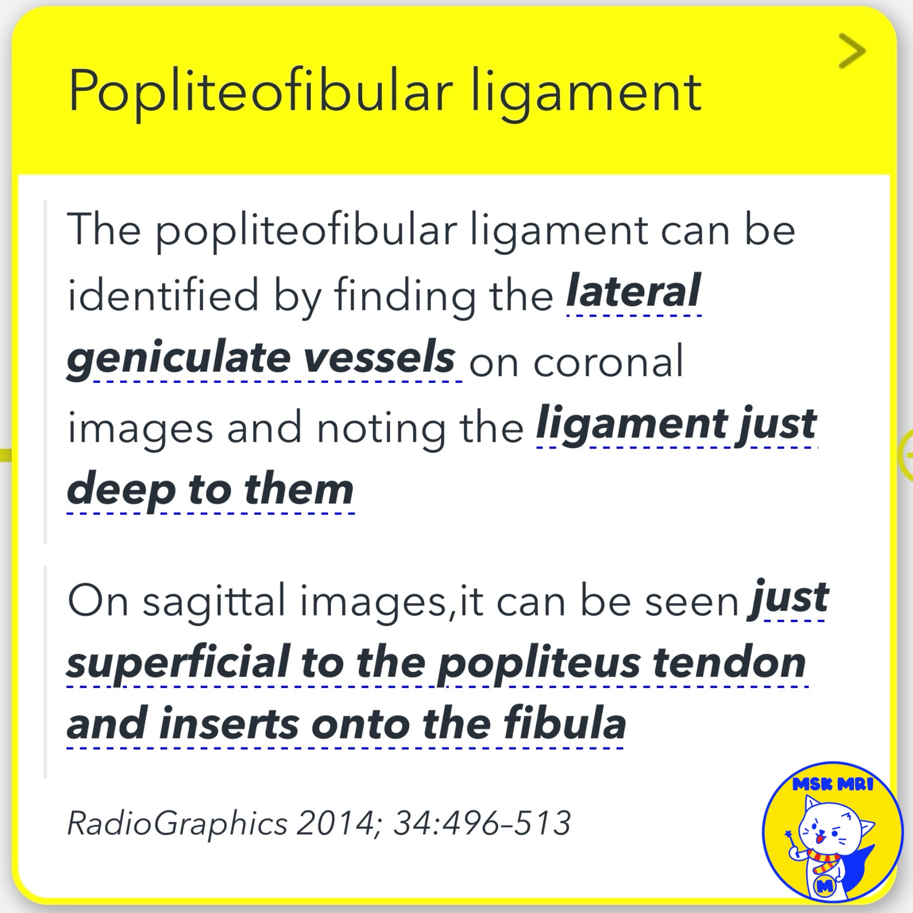
- On coronal images, it can be identified deep to the lateral geniculate vessels.
- On sagittal images, it is seen superficial to the popliteus tendon, inserting onto the fibula.
✅ Relationship with Anteroinferior Popliteomeniscal Fascicle
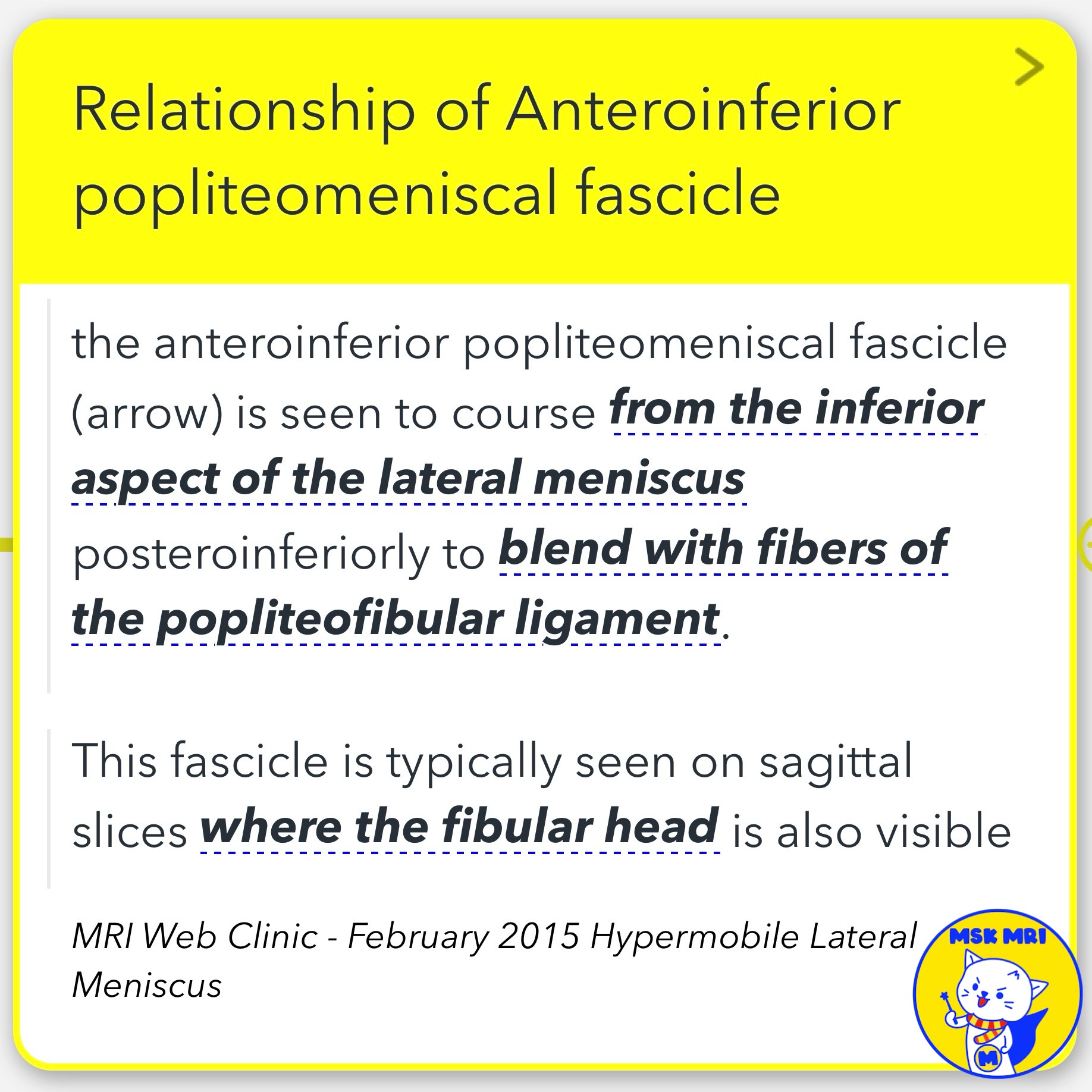
- Courses from the inferior aspect of the lateral meniscus posteroinferiorly.
- Blends with fibers of the popliteofibular ligament.
- Visible on sagittal slices showing the fibular head.
✅ Relationship with Arcuate Ligament
- More posterior and vertical than the popliteofibular ligament.
- Seen on slightly more medial sagittal sections.
- Has a more medial angle toward the posterior capsule.
RadioGraphics 2014; 34:496–513
MRI Web Clinic - February 2015 Hypermobile Lateral Meniscus
Radiol Clin North Am. 2018 Nov;56(6):935-951
"Visualizing MSK Radiology: A Practical Guide to Radiology Mastery"
© 2022 MSK MRI Jee Eun Lee All Rights Reserved.
No unauthorized reproduction, redistribution, or use for AI training.
#PopliteofibularLigament, #KneeAnatomy, #PosterolateralCorner, #TendinousBand, #LateralGeniculate, #PopliteusTendon, #FibularStyloid, #AnteroinferiorPopliteomeniscalFascicle, #ArcuateLigament, #PosteriorCapsule


'✅ Knee MRI Mastery > Chap 3.Collateral Ligaments' 카테고리의 다른 글
| (Fig 3-B.18) Popliteofibular Ligament Avulsion Fracture (0) | 2024.05.22 |
|---|---|
| (Fig 3-B.17) Partial Popliteofibular Ligament Tear (0) | 2024.05.22 |
| (Fig 3-B.15) Popliteus Tendon Avulsion Fracture (0) | 2024.05.21 |
| (Fig 3-B.14) Intra-Articular Partial Tear of the Popliteus (0) | 2024.05.21 |
| (Fig 3-B.13) Popliteus Myotendinous Junction Injury (0) | 2024.05.21 |