Click the link to purchase on Amazon 🎉📚
==============================================
🎥 Check Out All Videos at Once! 📺
👉 Visit Visualizing MSK Blog to explore a wide range of videos! 🩻
https://visualizingmsk.blogspot.com/?view=magazine
📚 You can also find them on MSK MRI Blog and Naver Blog! 📖
https://www.instagram.com/msk_mri/
Click now to stay updated with the latest content! 🔍✨
==============================================
📌The Popliteofibular Ligament
✅ Anatomy
- The popliteofibular ligament extends from the popliteus tendon near the myotendinous junction to the posteromedial aspect of the fibular styloid process, posteromedial to the biceps femoris tendon insertion.
- On coronal MRI images, the popliteofibular ligament can be identified just deep to the lateral geniculate vessels.
- Coronal images provide the best visualization of this ligament.
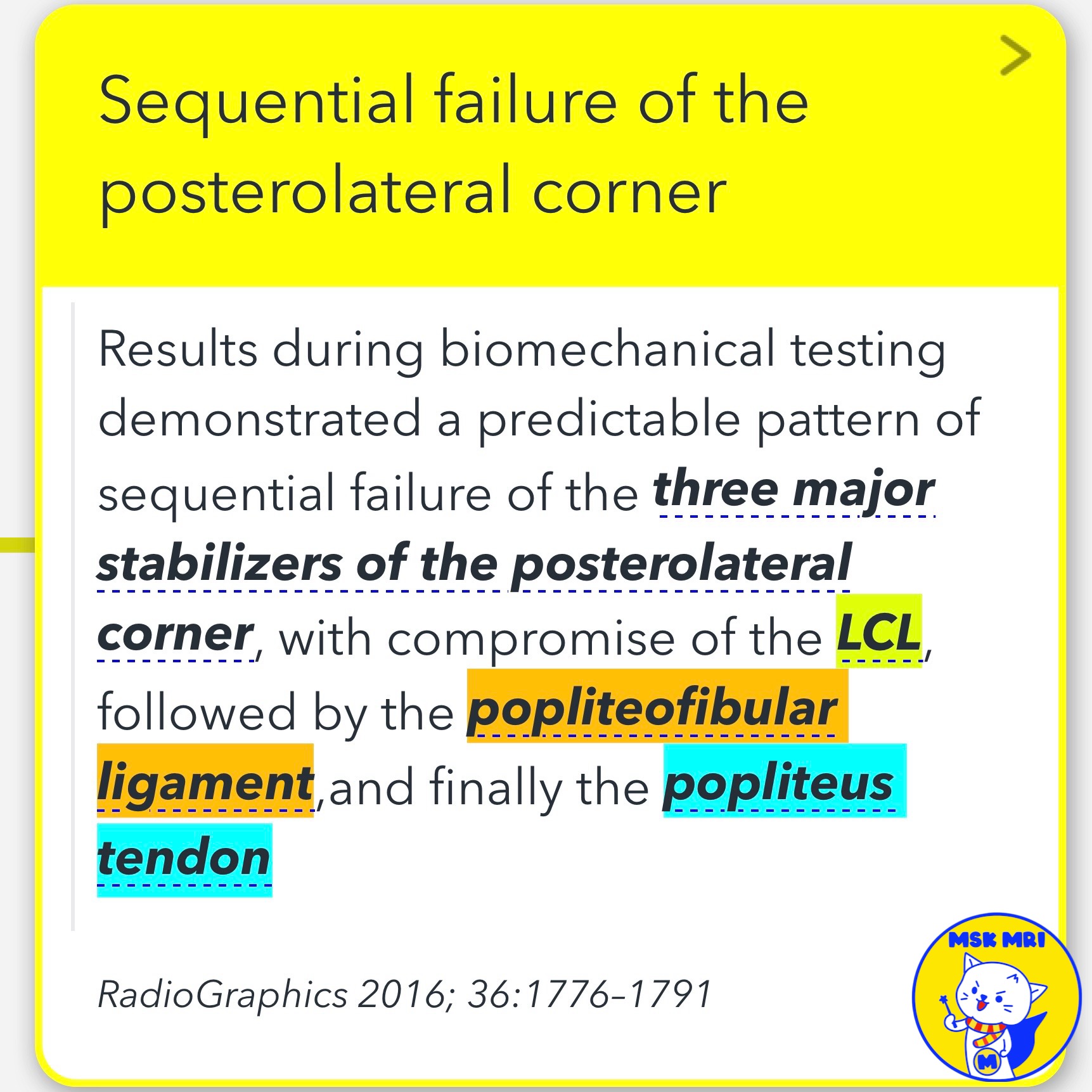
✅ Injury Patterns Avulsions
- Avulsions from the fibular styloid process can occur
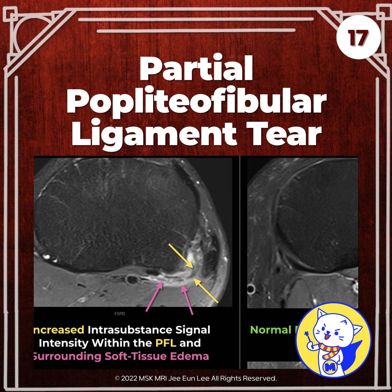
- Partial Tears: Typically appear as increased intrasubstance or peritendinous signal intensity on MRI
- Complete Tears: In the setting of a ruptured popliteofibular ligament, surgical reconstruction is currently advocated to restore normal tibiofemoral stability and kinematics.
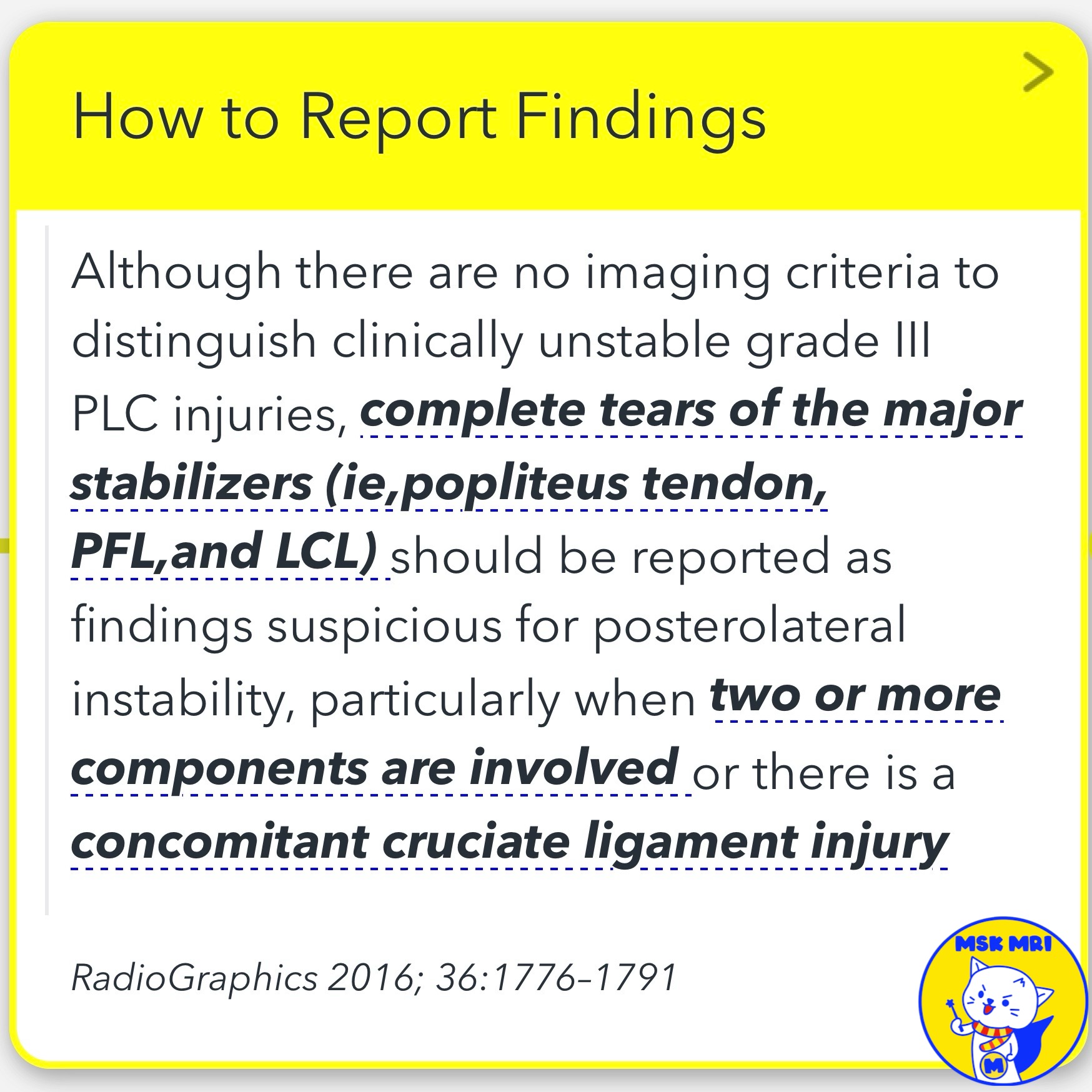
✅ Reporting Findings
- No imaging criteria to definitively diagnose posterolateral corner instability
- However, complete tears of the major stabilizers (popliteus tendon, popliteofibular ligament, and lateral collateral ligament) should be reported as suspicious for posterolateral instability, especially when : Two or more components are involved or There is a concomitant cruciate ligament injury
RadioGraphics 2016; 36:1776–1791
"Visualizing MSK Radiology: A Practical Guide to Radiology Mastery"
© 2022 MSK MRI Jee Eun Lee All Rights Reserved.
No unauthorized reproduction, redistribution, or use for AI training.
#popliteofibularligament, #posterolateralcornerinjury, #kneeinstability, #ligamenttear, #avulsioninjury, #MRIfindings, #orthopedicsurgery, #cruciateligamentinjury, #orthopedicimaging


'✅ Knee MRI Mastery > Chap 3.Collateral Ligaments' 카테고리의 다른 글
| (Fig 3-B.19) Fabellofibular Ligament Anatomy (0) | 2024.05.22 |
|---|---|
| (Fig 3-B.18) Popliteofibular Ligament Avulsion Fracture (0) | 2024.05.22 |
| (Fig 3-B.16) Popliteofibular Ligament Anatomy (0) | 2024.05.22 |
| (Fig 3-B.15) Popliteus Tendon Avulsion Fracture (0) | 2024.05.21 |
| (Fig 3-B.14) Intra-Articular Partial Tear of the Popliteus (0) | 2024.05.21 |