Click the link to purchase on Amazon 🎉📚
==============================================
🎥 Check Out All Videos at Once! 📺
👉 Visit Visualizing MSK Blog to explore a wide range of videos! 🩻
https://visualizingmsk.blogspot.com/?view=magazine
📚 You can also find them on MSK MRI Blog and Naver Blog! 📖
https://www.instagram.com/msk_mri/
Click now to stay updated with the latest content! 🔍✨
==============================================
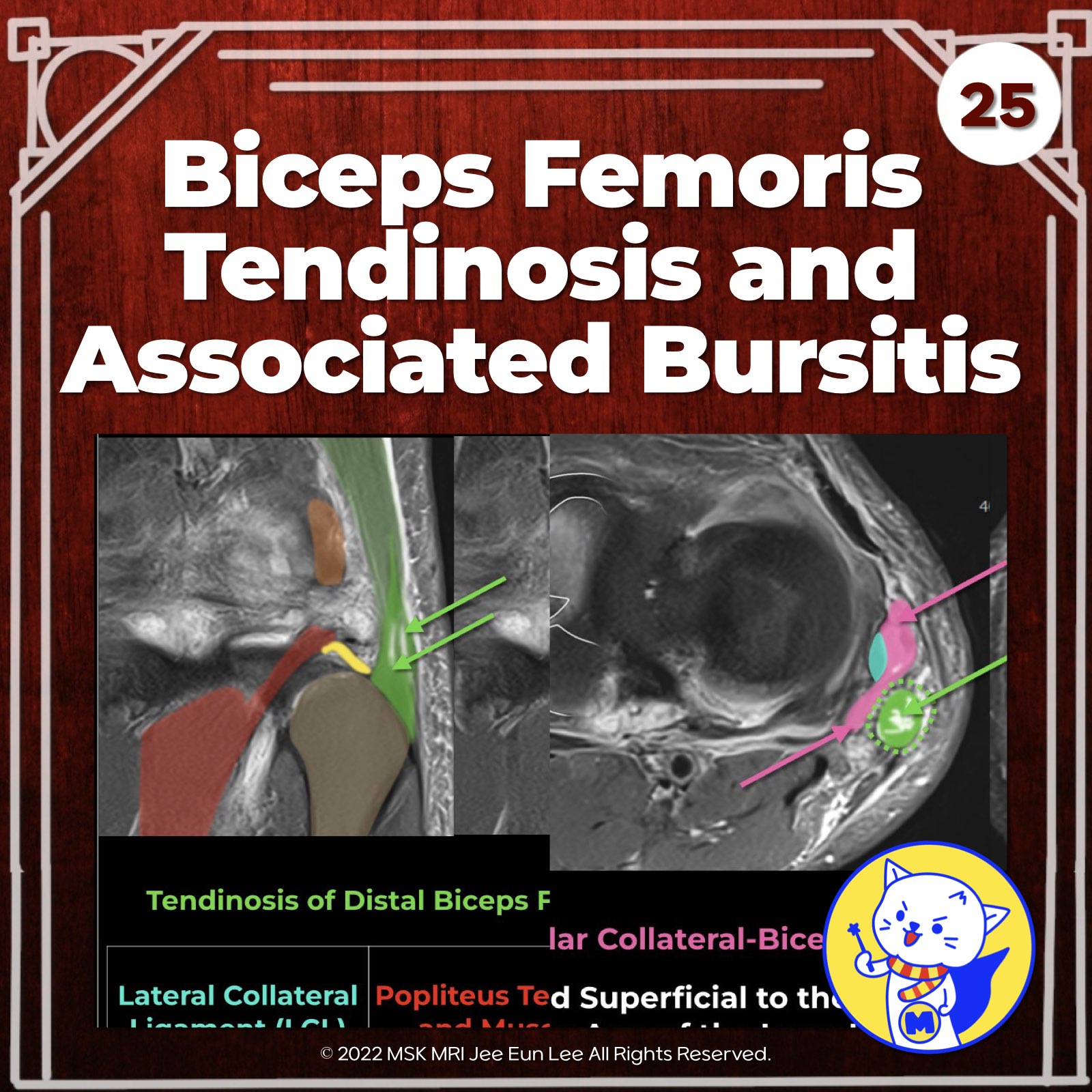
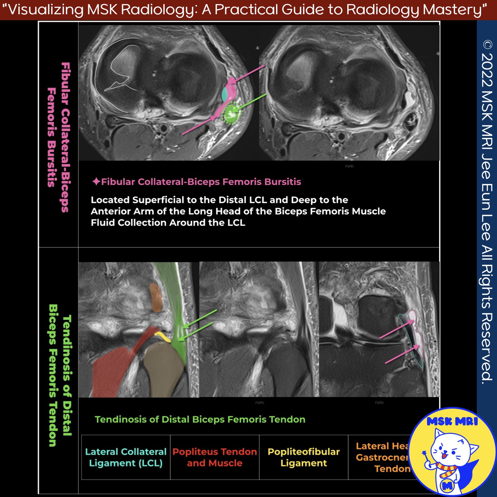
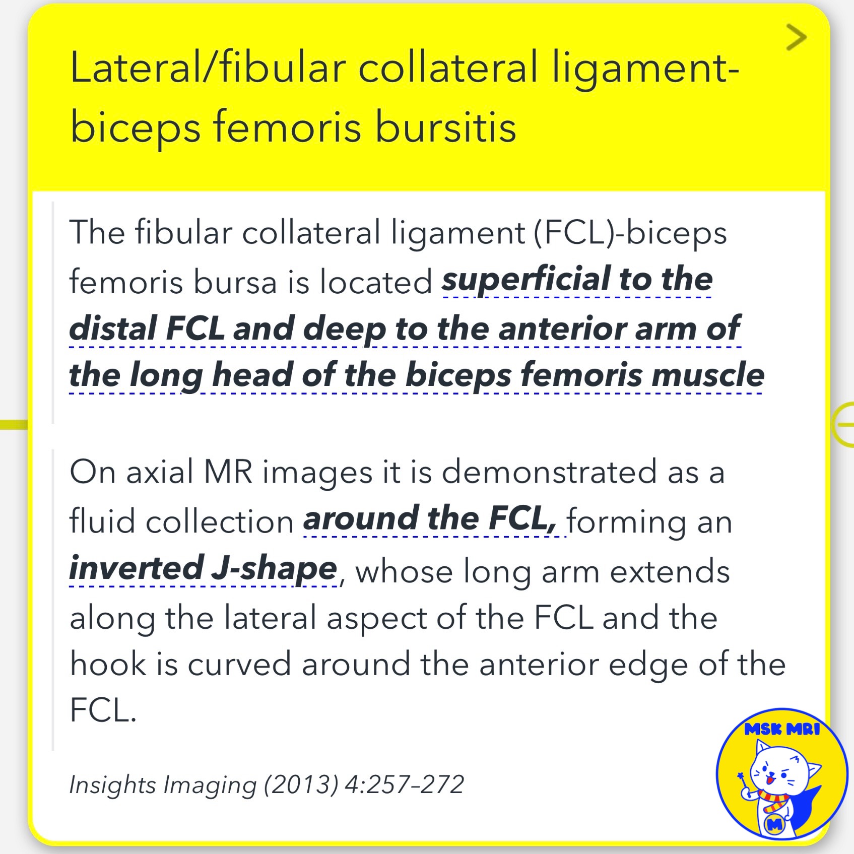
📌 Lateral/fibular collateral ligament-biceps femoris bursitis (LCL bursitis)
- The FCL-biceps femoris bursa is superficial to the distal FCL and deep to the biceps femoris anterior arm.
- On axial MRI, it has an inverted J-shape surrounding the FCL: the long arm along the lateral FCL aspect, the hook around the FCL's anterior edge.
- Proximally it reaches the biceps femoris anterior arm's superior margin, distally extending to the FCL's fibular insertion.
Insights Imaging (2013) 4:257–272
"Visualizing MSK Radiology: A Practical Guide to Radiology Mastery"
© 2022 MSK MRI Jee Eun Lee All Rights Reserved.
No unauthorized reproduction, redistribution, or use for AI training.
#FCLBursitis, #LateralCollateralLigamentBursitis, #BicepsFemorisBursitis, #KneeBursitis, #KneeAnatomyMRI,
#kneepain, #bicepsfemoristendinosis, #bursitis, #tendonthickening, #tendoninjuries,

'✅ Knee MRI Mastery > Chap 3.Collateral Ligaments' 카테고리의 다른 글
| (Fig 3-B.28) Proximal Anterolateral Ligament Tear (1) | 2024.05.23 |
|---|---|
| (Fig 3-B.26) Anterolateral Ligament Anatomy (0) | 2024.05.22 |
| (Fig 3-B.24) Distal Biceps Femoris Myotendinous Junction Tear (0) | 2024.05.22 |
| (Fig 3-B.23) Anatomy of Biceps Femoris Arms: Direct and Anterior (0) | 2024.05.22 |
| (Fig 3-B.22) Arcuate and Fabellofibular Ligament Tears (0) | 2024.05.22 |