https://youtu.be/vzxclU_34OI?si=B88NykUJHSELzyAC
Click the link to purchase on Amazon 🎉📚
==============================================
🎥 Check Out All Videos at Once! 📺
👉 Visit Visualizing MSK Blog to explore a wide range of videos! 🩻
https://visualizingmsk.blogspot.com/?view=magazine
📚 You can also find them on MSK MRI Blog and Naver Blog! 📖
https://www.instagram.com/msk_mri/
Click now to stay updated with the latest content! 🔍✨
==============================================
📌 Relationship with the Superficial MCL
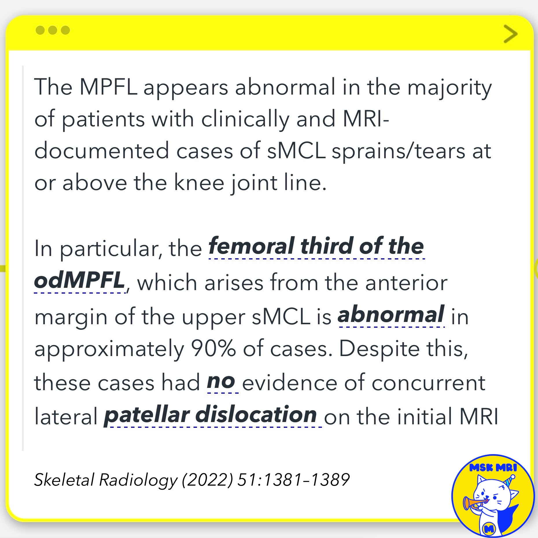
- The sMCL attaches proximal and posterior to the medial epicondyle. In cases of sMCL tears, the femoral third of the oblique decussation MPFL component (arising from the anterior upper sMCL) is abnormal in approximately 90% of cases, despite no evidence of lateral patellar dislocation on initial MRI.
✅ The Oblique Decussation Component of the MPFL
- Recently described broader portion of the MPFL arising from anterior margin of upper superficial MCL
- MRI shows abnormal appearance in ~90% of MCL sprains/tears
- Despite abnormality, no concurrent lateral patellar dislocation seen initially on MRI
- Reflects intimate anatomic relationship and shared fibers between odMPFL and MCL
- Abnormal odMPFL does not necessarily indicate patellar instability based on MRI alone
★ Key Takeaway: Radiologists should recognize odMPFL can appear abnormal with MCL injuries, but this does not definitively diagnose patellar dislocation.
Radiographics. 2023 Jun;43(6):e220177
Skeletal Radiology (2022) 51:1381–1389
"Visualizing MSK Radiology: A Practical Guide to Radiology Mastery"
© 2022 MSK MRI Jee Eun Lee All Rights Reserved.
No unauthorized reproduction, redistribution, or use for AI training.
#MPFL, #AdductorTubercle, #KneeLigament, #sMCL, #KneeMRI, #FemoralAttachment, #KneeAnatomy, #Orthopedics, #LigamentInjury, #MedicalImaging
'✅ Knee MRI Mastery > Chap 4A. Patelloefemoral joint' 카테고리의 다른 글
| (Fig 4-A.41) Chronic Patellofemoral Instability: Part 2 (0) | 2024.06.06 |
|---|---|
| (Fig 4-A.40) Chronic Patellofemoral Instability: Part 1 (0) | 2024.06.06 |
| (Fig 4-A.38) Vastus medialis oblique (VMO) Muscle Injury (0) | 2024.06.06 |
| (Fig 4-A.37) Tear of the Medial Retinaculum and Patellar Tendon (0) | 2024.06.06 |
| (Fig 4-A.36) Complete Tear of the Medial Retinaculum (0) | 2024.06.06 |


