https://youtu.be/vzxclU_34OI?si=B88NykUJHSELzyAC
Click the link to purchase on Amazon 🎉📚
==============================================
🎥 Check Out All Videos at Once! 📺
👉 Visit Visualizing MSK Blog to explore a wide range of videos! 🩻
https://visualizingmsk.blogspot.com/?view=magazine
📚 You can also find them on MSK MRI Blog and Naver Blog! 📖
https://www.instagram.com/msk_mri/
Click now to stay updated with the latest content! 🔍✨
==============================================
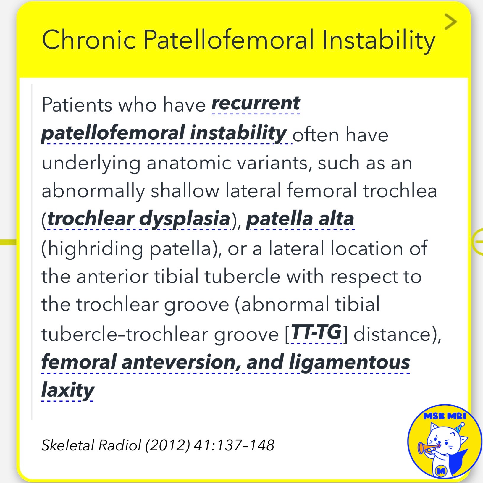
📌 Chronic Patellofemoral Instability
✅ Manifestations of Chronic Patellofemoral Instability
- Fibrosed/thickened/attenuated retinaculum
- Bony productive changes at retinacular attachments
- Ossification of retinacular layers
- Asymmetric retinacular thickening/attenuation
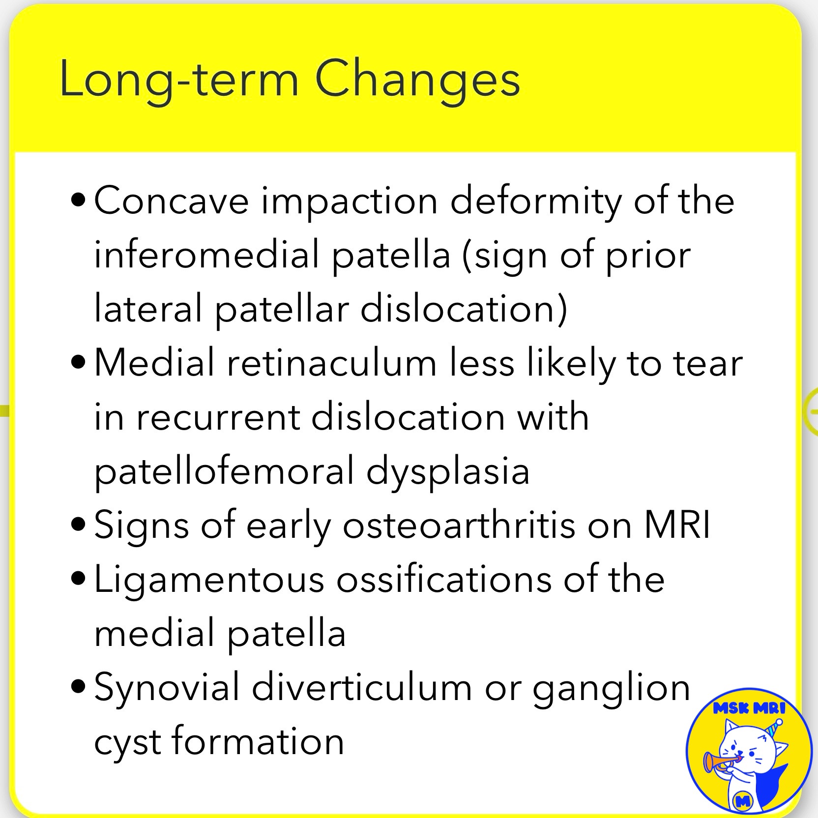
✅ Long-term Changes
- Signs of early osteoarthritis on MRI
- Ligamentous ossifications of the medial patella
- Synovial diverticulum or ganglion cyst formation
✅ Specific Findings
- Concave impaction deformity of the inferomedial patella (sign of prior lateral patellar dislocation)
- Medial retinaculum less likely to tear in recurrent dislocation with patellofemoral dysplasia
✅ Predisposing Factors
- Trochlear dysplasia (abnormally shallow lateral femoral trochlea)
- Patella alta (high-riding patella)
- Abnormal tibial tubercle–trochlear groove (TT-TG) distance
- Femoral anteversion
- Ligamentous laxity
References:
Skeletal Radiol (2012) 41:137–148
Radiographics. 2010 Jul-Aug;30(4):961-81
Radiology. 2002 Dec;225(3):736-43
Radiol Clin N Am 51 (2013) 393–411
#ChronicPatellofemorialInstability, #OsteoarthritisChanges, #LigamentousOssifications, #SynovialDiverticulum, #ConcaveImpactionDeformity, #TrochlearDysplasia, #PatellaAlta, #AbnormalTT-TGDistance, #FemoralAnteversion, #LigamentousLaxity


'✅ Knee MRI Mastery > Chap 4A. Patelloefemoral joint' 카테고리의 다른 글
| (Fig 4-A.42) MPFL Reconstruction and Lateral Release (0) | 2024.06.07 |
|---|---|
| (Fig 4-A.41) Chronic Patellofemoral Instability: Part 2 (0) | 2024.06.06 |
| (Fig 4-A.39) Medial Patellofemoral Ligament Abnormality with MCL Injury (0) | 2024.06.06 |
| (Fig 4-A.38) Vastus medialis oblique (VMO) Muscle Injury (0) | 2024.06.06 |
| (Fig 4-A.37) Tear of the Medial Retinaculum and Patellar Tendon (0) | 2024.06.06 |