Wrist, Finger MRI 35

[Anatomy_21] Physiologic displacement of the extensor carpi ulnaris Tendon, Pronation, Supination

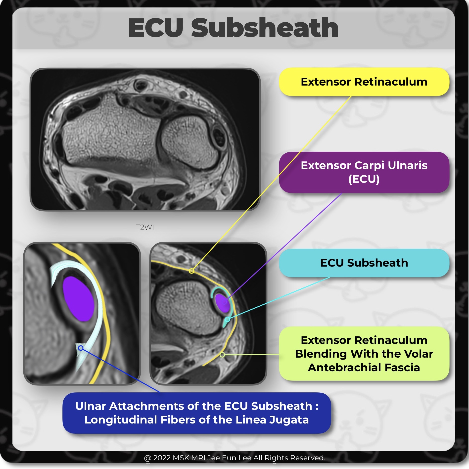
In the neutral and pronation positions, the ECU tendon is centered in the groove in the distal portion of the ulna. Note the normal tendency of the ECU tendon to sublux to the apex of the ulnar styloid at the ulnar border of the groove during supination. Variations in length of distal extension of the ECU subsheath connecting the ulnar styloid process and superficial ligamentous fibers of the do..

[Anatomy_20] Extensor carpi ulnaris (ECU) subsheath complex, Linea Jugata

ECU subsheath connected the ulnar styloid process and superficial ligamentous fibers of the dorsal radioulnar ligament. So, The subsheath of the ECU tendon maintains the stability of the dorsal aspect of the TFCC it is necessary to learn about the ECU subsheath. The extensor retinaculum is an important and complex structure, preventing extensor tendons bowstringing. The extensor retinaculum cour..

[Anatomy_19] Distal Insertion of the ulnomeniscal homologue, TFCC, wrist MRI

The fibers at the main insertion into the ulnar aspect of the triquetrum are recognized between the triquetrum and the ECU tendon. Some fibers attach to the ulnar aspect of the pisiform (P), forming the dorsal capsular ligament of the pisotriquetral joint. The entire distal insertion was clearly separated from the volar extrinsic ligaments. #WristMRI, #WristAnatomy, #WristLigament, #WristInstabi..

[Anatomy_18] Component of ulnomeniscal homologue, TFCC, wrist MRI

(Styloid component) this component is recognized between the entrance to the prestyloid recess and the the extensor carpi ulnaris tendon (ECU). (Collateral component), UMH, the ECU tendon sheath, and the joint capsule (ulnar collateral ligament) is fused indistinguishably. (Distal insertion) The ulnar aspect of the triquetrum represented the main insertion site. #WristMRI, #WristAnatomy, #WristL..

[Anatomy_17] Ulnomeniscal homologue, Radioulnar Ligament TFCC, Ulnotriquetral Ligament, Ulnotriquetral

The ulnomeniscal homologue (UMH) is noted slightly ulnar to the origin of the dorsal radioulnar ligament and radioulnar component of the UMH. The radioulnar component of the UMH arose at the dorsal radioulnar ligament of the TFC complex. The distal insertion of the UMH is seen with its main body attaching to the triquetrum #WristMRI, #WristAnatomy, #WristLigament, #WristInstability, #TFCC, #Wris..

[Anatomy_16] Radioulnar Component of ulnomeniscal homologue, TFCC, wrist MRI

At the level of the origin of the dorsal radioulnar ligament, radioulnar component of the ulnomeniscal homologue (UMH) is noted. The radioulnar component of the UMH arose at the dorsal edge of the radial notch, together with the dorsal radioulnar ligament of the TFC complex. The broad ligamentous structure attaches to the dorsal edge of the radial notch and divides into the proximal and distal l..

[Anatomy_15] Triangular fibrocartilage complex (TFCC), wrist MRI

The volar and dorsal aspects of the attachment correspond to slip insertions of the radioulnar ligaments into bone. The central portion of the attachment appears as an insertion into the hyaline cartilage of the radius The triangular fibrocartilage or articular disc of the TFCC has a broad attachment to the hyaline cartilage of the sigmoid notch of the distal radius, whereas, the dorsal and vola..

[Anatomy_14] Triangular fibrocartilage complex (TFCC), wrist MRI

On the ulnar attachment, TFCC attaches onto the ulnar styloid process through the distal lamina and onto the fovea of distal ulna through the proximal lamina; they are separated by the ligamentum subcruentum. The most important structure of the TFCC is the triangular fibrocartilage (TFC) proper, a triangular-shaped piece of fibrocartilage. On the radial side, the triangular fibrocartilage or art..

[Anatomy_13] Ulnotriquetral Ligament, Two Normal Perforation, prestyloid recess, pisotriquetral joint orifice

The ulnotriquetral ligament (UT) often has 2 normal perforations: a proximal opening to the prestyloid recess, and a distal pisotriquetral joint orifice. (Left image) The prestyloid recess perforation is found just ulnar to the union between the proximal radioulnar ligament and the UT ligament. (Right image) The pisotriquetral orifice is anterior to the proximal articular surface of the triquetr..

[Anatomy_12] Ulnotriquetral Ligament, TFCC, wrist MRI

The ulnotriquetral ligament (UT) forms the anterior and ulnar part of the ulnocarpal joint capsule together with the ulnolunate ligament. UT shares a common origin and connects the volar radioulnar ligament and thus the triangular fibrocartilage complex to the triquetral bone. #WristMRI, #WristAnatomy, #WristLigament, #WristInstability, #TFCC, #Wrist MRI, #Wrist anatomy, #wrist ligament, #안산에이스병..