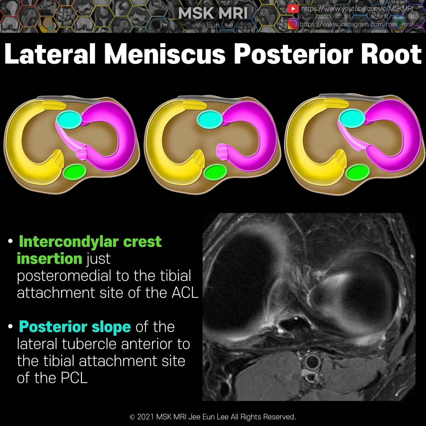
PRLM has two attachment sites of the intertubercular region and the posterior slope of the lateral tubercle. Contiguous fat-suppressed PD coronal images with a posteroanterior direction demonstrate small, dark signal foci (white arrows) traversing the intertubercular crest and terminating on the medial tubercle, near the tibial attachment of the ACL. These green-yellow arrows show an intercondyl..