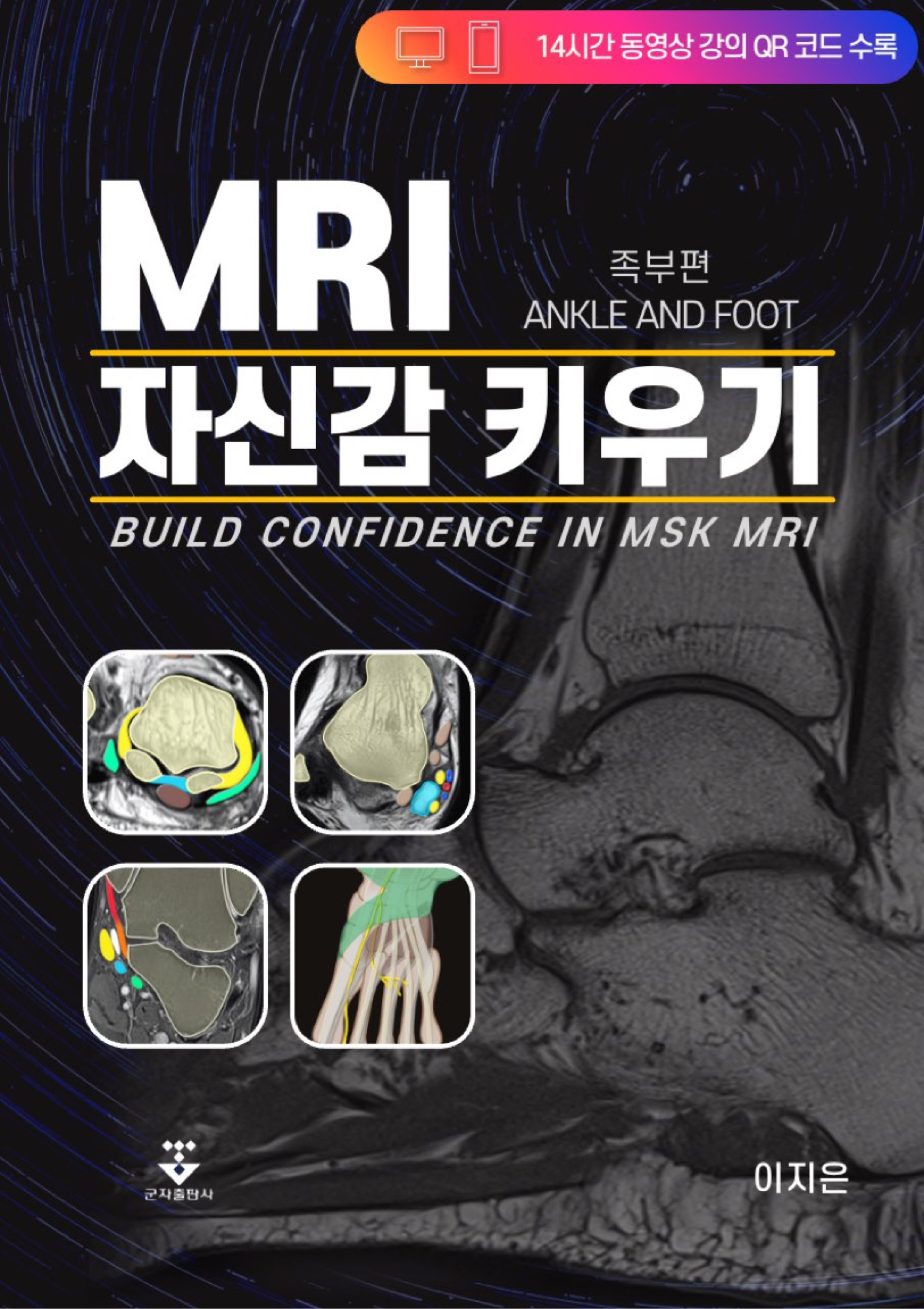
MSKMRI 39

[MRI ์ž์‹ ๊ฐํ‚ค์šฐ๊ธฐ-02] Deltoid ligament, Ankle MRI anatomy, tibiotalar ligament, ๊ตฐ์ž์ถœํŒ์‚ฌ

Ankle MRI, Anatomy, ankle ligament, ๊ตฐ์ž์ถœํŒ์‚ฌ, MRI์ž์‹ ๊ฐ ํ‚ค์šฐ๊ธฐ, MSKMRI, deltoid ligament, posterior tibiotalar ligament, tibiospring ligament, superficial layer, deep layer, tibiocalcaneal ligament, tibionavicular ligament https://cafe.naver.com/studywithmskmri/361 [MRI ์ž์‹ ๊ฐํ‚ค์šฐ๊ธฐ-02] Deltoid ligament, Ankle MRI anatomy, tibiotalar ligament, ๊ตฐ์ž์ถœํŒ์‚ฌ Ankle MRI, Anatomy, ankle ligament, ๊ตฐ์ž์ถœํŒ์‚ฌ, MRI์ž์‹ ๊ฐ ํ‚ค์šฐ๊ธฐ, MSKMRI..

[HYPERLINK] Athletic pubalgia, core muscle injuries, rectus abdominis, adductor longus aponeurosis, pubic plate injury, secondary cleft sign (SITE MAP)

Athletic pubalgia, core muscle injuries, rectus abdominis, adductor longus aponeurosis, pubic plate injury, secondary cleft sign 01 Anatomy, Athletic pubalgia, core muscle injuries, rectus abdominis, adductor longus aponeurosisโ€‹ 02 Rectus Abdominis Adductor Aponeurosis Injury Athletic pubalgia, core muscle injuries 03 Midline aponeurotic pubic plate injury, secondary cleft sign, Athletic pubalgi..

[HYPERLINK] Shoulder MRI checklist (SITE MAP)

Shoulder MRI checklist โ€‹ [Shoulder_01] MRI checklist, Coronal image, rotator cuff, SLAP lesion, calcific tendinitis, biceps โ€‹ [Shoulder_02] MRI checklist, Axial image, AC joint, SLAP, Hill-Sachs, Bankart, rotator cuff tear, โ€‹ [Shoulder_03] MRI checklist, Sagittal image, rotator cuff tear, acromion, biceps tendon, adhesive (์นดํŽ˜์— ๊ฐ€์ž…ํ•˜์…”์•ผ ๋‚˜๋จธ์ง€ ๋™์˜์ƒ ๊ฐ•์˜๋ฅผ ๋ณด์‹ค ์ˆ˜ ์žˆ์Šต๋‹ˆ๋‹ค.) ์Šคํ„ฐ๋””์œ„๋“œ MSKMRI : ๋„ค์ด๋ฒ„ ์นดํŽ˜ MSK MRI์™€ ์ฆ๊ฒ๊ฒŒ ๊ณต๋ถ€ํ•˜๊ณ ..

[MRI ์ž์‹ ๊ฐ ํ‚ค์šฐ๊ธฐ: ์กฑ๋ถ€ํŽธ] ์ œ ์ฑ…์ด ์ถœ๊ฐ„๋˜์—ˆ์Šต๋‹ˆ๋‹ค.

์•ˆ๋…•ํ•˜์„ธ์š” ์˜์ƒ์˜ํ•™๊ณผ ์ด์ง€์€์ž…๋‹ˆ๋‹ค. 2020๋…„ ๊ฒจ์šธ๋ถ€ํ„ฐ ์“ฐ๊ธฐ ์‹œ์ž‘ํ•œ ์ œ ์ฑ…์ด ๋“œ๋””์–ด ์ถœ๊ฐ„๋˜์—ˆ์Šต๋‹ˆ๋‹ค. ์ด๋ฏธ์ง€๊ฐ€ ์›Œ๋‚™ ๋งŽ๊ณ , ๋™์˜์ƒ QR์ฝ”๋“œ๊ฐ€ ์ˆ˜ ๋ฐฑ๊ฐœ์ด๋‹ค๋ณด๋‹ˆ 1๋…„์ด ๋„˜๊ฒŒ ๊ฑธ๋ ธ๋„ค์š”. (๊ตฐ์ž์ถœํŒ์‚ฌ์™€ ์ œ๊ฐ€ 13์ฐจ ์ˆ˜์ •๊นŒ์ง€ ํ–ˆ๋”๋ผ๊ตฌ์š”) ์ œ ์ฑ…์„ ๊ธฐ๋‹ค๋ ค ์ฃผ์‹  ์„ ์ƒ๋‹˜๋“ค์—๊ฒŒ ์ƒ๊ฐ๋ณด๋‹ค ์˜ค๋ž˜ ๊ฑธ๋ ค์„œ ์ฃ„์†กํ•˜๋‹ค๋Š” ๋ง์”€์„ ๋“œ๋ฆฌ๊ณ  ์‹ถ์Šต๋‹ˆ๋‹ค. ์ œ ์ฑ…์˜ ์—ฌ๋Ÿฌ ํŒ๋งค์ฒ˜ ์ค‘ ๋ช‡ ๊ตฐ๋ฐ๋งŒ ๋งํฌ๋ฅผ ์•„๋ž˜ ๋‹ฌ์•„๋‘๊ฒ ์Šต๋‹ˆ๋‹ค. ๋ถ€์กฑํ•  ์ˆ˜ ์žˆ์ง€๋งŒ, ์ตœ์„ ์„ ๋‹คํ•œ ์ฑ…์ด๋‹ˆ ๋งŽ์€ ๊ด€์‹ฌ ๊ฐ€์ ธ์ฃผ์‹œ๋ฉด ๊ฐ์‚ฌ๋“œ๋ฆฌ๊ฒ ์Šต๋‹ˆ๋‹ค ^^ MRI ์ž์‹ ๊ฐ ํ‚ค์šฐ๊ธฐ: ์กฑ๋ถ€ํŽธ ๋„ค์ด๋ฒ„ ์ฑ…: MRI ์ž์‹ ๊ฐ ํ‚ค์šฐ๊ธฐ 2020๋…„ ๋ด„, ๊ฐœ์ธ ์œ ํŠœ๋ธŒ ์ฑ„๋„์„ ํ†ตํ•ด ์ •๋ฆฌํ•˜๋ฉฐ ๊ณต๋ถ€ํ•˜๋˜ ๊ฒƒ์„ ๋™๋ฃŒ, ํ›„๋ฐฐ ์„ ์ƒ๋‹˜๋“ค๊ป˜ ๋„ ์›€์ด ๋˜๋Š” ๋ถ€๋„๋Ÿฝ์ง€ ์•Š์€ ์ฑ…์œผ๋กœ ๋งŒ๋“ค์–ด๋ณด์ž๋Š” ์ƒ๊ฐ์— ๊ณ„์†๋œ ์ˆ˜์ •๊ณผ ๋‚ด์šฉ ์ถ”๊ฐ€๋ฅผ ๋ฐ˜๋ณตํ•˜ ๋‹ค๋ณด๋‹ˆ..

[Anatomy_31] Anterior meniscofemoral ligament, โ€‹โ€‹Anomalous insertion of the medi

Anomalous insertion of the medial meniscus (AIMM) into the anterior cruciate ligament (ACL) is rare.The AIMM has the same anatomical description as the anteromedial meniscofemoral ligament It appears as a linear band isointense to the ACL and meniscus, which runs parallel to the ACL and demonstrates a hyperintense gap between it and the ACL On MRI, this anomaly may be misinterpreted as an ACL te..

[Anatomy_30] Oblique Meniscomeniscal Ligament vs bucket handle tear -03

The medial OMML originates from the anterior horn of the medial meniscus and inserts onto the posterior horn of the lateral meniscus between the ACL and PCL. This structure should not be mistaken for a bucket handle meniscal tear A bucket-handle tear is a longitudinal tear with central migration of the inner “handle” fragment The correct diagnosis of an oblique meniscomeniscal ligament rather th..

Knee MRI/Meniscus 2021.10.02