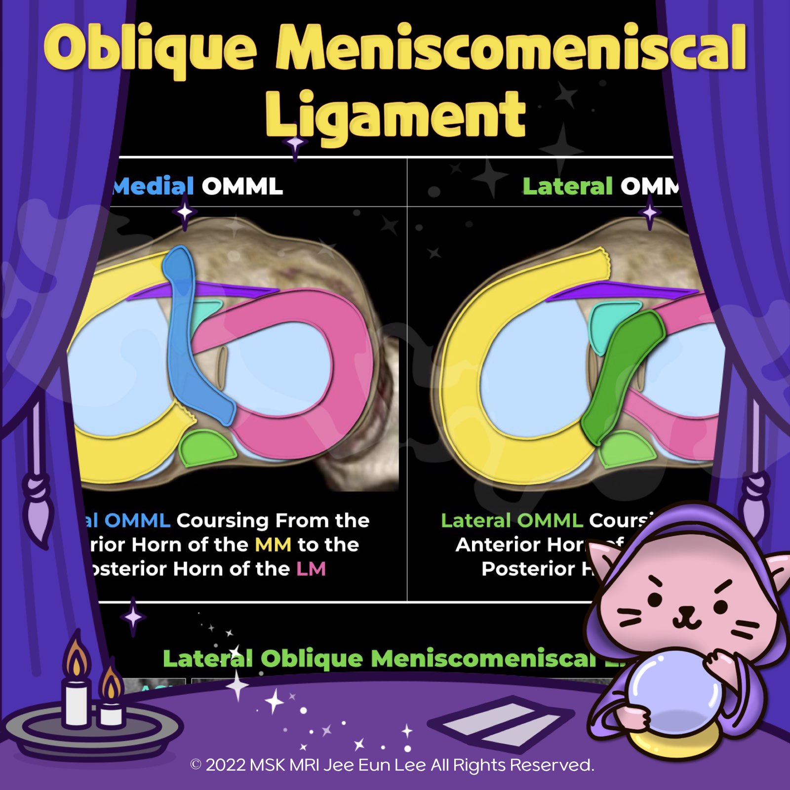
MSKMRI 39

(Fig 1-A.15) Additional peripheral attachments to the MM

https://youtu.be/C-I1T1POols https://youtu.be/kEuMzUrHD0M The knee joint capsule, part of layer III, forms the deep MCL, with its meniscotibial and meniscofemoral components, along the medial aspect of the knee. It then continues posteriorly, where it is reinforced externally by the POL and expansions from the semimembranosus. More posteriorly, the posteromedial joint capsule passes deep to the ..

(Fig 1-A.14) anatomy of the Posterior Medial Capsule (PMC)

https://youtu.be/H1BeOmollx4 https://youtu.be/kEuMzUrHD0M Component Description Attachments The meniscocapsular ligament extends to the capsule; The meniscotibial ligament attaches to the tibia. Tear Patterns Five patterns of ramp injury; common types are full-thickness tears at the capsulosynovial junction and in the red zone. Fat A thin fat pad separates the ramp capsule and meniscotibial liga..

(Fig 1-A.13) normal popliteomeniscal fascicle deficiency

https://youtu.be/ckDHFabBkUk?si=01vgw2dU-6veGl1S https://youtu.be/kEuMzUrHD0M 👉Anteroinferior Popliteomeniscal Fascicle: This fascicle forms the lateral floor of the popliteal hiatus, varying in thickness. It connects the lateral meniscus’s body, extends to the popliteus muscle’s musculotendinous unit, and blends with the popliteofibular ligament, inserting at the fibular head’s styloid process...

(Fig 1-A.06) Medial Meniscus Posterior Root, Anatomy, degeneration, tear

https://youtu.be/CjYENTgOT3Y https://youtu.be/kEuMzUrHD0M "🧐 Deep Dive into the Meniscofemoral Ligament (MFL) Anatomy! ✨ The MFL, a critical component of the knee, originates from the posterior horn of the lateral meniscus and inserts onto the lateral aspect of the posterior medial femoral condyle. It plays a key role in the stability and function of the knee joint. 🔍 Let's break it down: 1️⃣ Th..

(Fig 1-A.05) Anatomy of the Lateral Meniscus Posterior Root, two components, intertubercular area

https://youtu.be/-lNJo4zWnqs https://youtu.be/kEuMzUrHD0M PRLM has two attachment sites of the posterior slope of the lateral tubercle (1st coronal image) and intertubercular region (other coronal images). - major component: intertubercular area, anterior extension into medial tubercle along the intertubercular crest, just posteromedial to tibial attachment of anterior cruciate ligament (; ACL) ..

(Fig 1-A.06) Medial Meniscus Posterior Root, Anatomy, degeneration, tear

https://youtu.be/GvR9ULGrDOw https://youtu.be/kEuMzUrHD0M Category Description Insertion Points - Inserts along the downward slope of the intercondylar fossa, anterior to the posterior cruciate ligament. - Extends from the tibial attachment point to just lateral from the articular cartilage inflection point of the medial tibial plateau. - Commences just lateral to the apex of the medial tibial p..

(Fig 1-A.04) Anatomy of lateral Meniscus anterior root, tibial spine fracture

https://youtu.be/w2QemJqJBfsTitle: Unraveling the Complexities of the Lateral Meniscus and ACL on MRI https://youtu.be/kEuMzUrHD0M 🔍 Deep Dive into Anatomy and Injury: the intricate relationship between the anterior root of the lateral meniscus (LM) and the anterior cruciate ligament (ACL) in the knee, as revealed by MRI. Unique Structure: The anterior root of the LM is distinct with its fan-sha..

Anatomy of Medial Meniscus anterior root, variation

https://youtu.be/dECIy2gvacM https://youtu.be/kEuMzUrHD0M The medial meniscus has an anterior root that attaches to the area in front of the tibial attachment of the ACL, specifically in the intercondylar fossa of the tibia. Additionally, the posterior fibers of the anterior horn of the medial meniscus connect to the transverse ligament. This anterior root can vary in attachment, with most peopl..

[PCL 07] 02. Popliteus musculotendinous complex, popliteomeniscal fascicle, popliteal bursa, cyamella

With popliteus tears, it is important to note - the site of the tear (intra- articular or extra-articular), - the extent of tear (partial or full-thickness), - the presence of a bone fragment if there has been tendon avulsion - degree of tendon retraction Partial-thickness tear of the popliteus tendon, High-grade partial tearing of the PT at its femoral insertion Most popliteus tears are extra-a..

Knee MRI 2022.11.19