
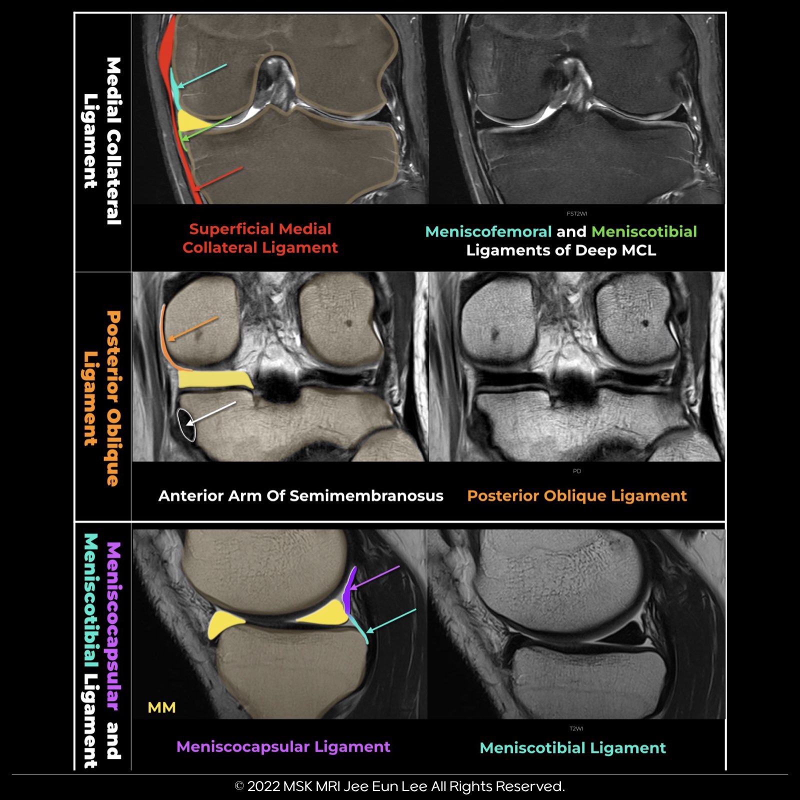

The knee joint capsule, part of layer III, forms the deep MCL, with its meniscotibial and meniscofemoral components, along the medial aspect of the knee. It then continues posteriorly, where it is reinforced externally by the POL and expansions from the semimembranosus.
More posteriorly, the posteromedial joint capsule passes deep to the medial head of the gastrocnemius. The two peripheral attachments of the MM’s posterior horn are the meniscocapsular ligament, extending posterosuperiorly to the capsule, and the meniscotibial ligament, which attaches inferiorly to the tibia.
Additional attachments of the MM: “Several structures attach to the medial meniscus (MM), serving as secondary stabilizers of the knee. These include the deep medial collateral ligament (MCL), posterior oblique ligament (POL), and semimembranosus tendon, among others.”
dMCL
The dMCL connects the meniscus to the femur and the tibia in the medial part of the knee and is a thickening of the medial joint capsule (layer 3)
It extends distally from posterior to anterior and terminates in a fan-like wide attachment on the tibia. The dMCL had firm meniscofemoral and meniscotibial attachment to the midbody of the medial meniscus, located between the meniscofemoral attachment of the POL and the anteromedial capsule.
POL
It then continues posteriorly, where it is reinforced externally by the POL and expansions from the semimembranosus The three arms of the POL are the superficial, central, and capsular arms. The distal insertions of the POL include the posteromedial capsule, SMT sheath, PHMM, and proximal tibia. The central arm was the largest and thickest portion of the posterior oblique ligament Its distal attachment was primarily to the posteromedial aspect of the medial meniscus, the meniscotibial portion of the posteromedial capsule, and the posteromedial part of the tibia The POL is part of layer II, and its anterior margin blends with the posterior margin of the superficial MCL Posteriorly, it blends with the joint capsule (layer III) and is inseparable from it
semimembranosus
“This semimembranosus-meniscal attachment originates from the anterior arm of the semimembranosus and is situated between the posterior meniscotibial ligament and the meniscotibial attachments of the posterior oblique ligament (POL).” “Some authors refer to the short meniscus arm of the semimembranosus. This arm attaches to the coronary ligament at the posterior horn of the medial meniscus.”
© 2022 MSK MRI Jee Eun Lee All Rights Reserved. You may not distribute or commercially exploit the content. Nor may you transmit it or store it on any other website or other forms of the electronic retrieval system. If you would like to use an image or video for anything other than personal use, please contact me. (jamaisvu1977@gmail.com), (jamaisvu77@naver.com) or instagram (msk_mri)
'✅ Knee MRI Mastery > Chap 1. Meniscus' 카테고리의 다른 글
| (Fig 1-A.17) Oblique meniscomeniscal ligament (0) | 2024.01.15 |
|---|---|
| (Fig 1-A.16) Anterior intermeniscal ligament (0) | 2024.01.14 |
| (Fig 1-A.14) anatomy of the Posterior Medial Capsule (PMC) (0) | 2024.01.12 |
| (Fig 1-A.13) normal popliteomeniscal fascicle deficiency (2) | 2024.01.11 |
| (Fig 1-A.12) Anatomy of popliteomeniscal fascicles (0) | 2024.01.10 |