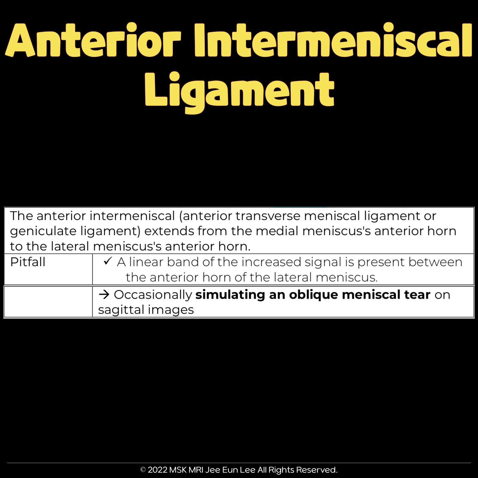
“The anterior intermeniscal ligament extends between the anterior horns of the medial and lateral menisci.
This structure is known by various names: the anterior inter-meniscal ligament (AIML), transverse meniscal ligament, ligamentum transversum genus, menisco-meniscal ligament, anterior transverse ligament, or inter-meniscal ligament. Its reported prevalence ranges from 3.6% to 100%.
A high-signal-intensity line is often observed in the lateral meniscus (LM), at the ligament’s attachment to the anterior horn’s superior surface. This line could be misinterpreted as an anterior horn tear.” This structure behind ARMM is an anterior intermeniscal ligament, so you can see that the posterior fibers of this medial meniscus anterior horn are stuck like this because the Posterior fibers of the MM anterior horn attach to the transverse ligament. This finding is normal.
An absent anterior root medial meniscus attachment is reported in 3% of individuals; the medial meniscus is instead stabilized by the anterior inter-meniscal ligament (AIML)
At the attachment of the ligament onto the superior surface of the anterior horn of the lateral meniscus, there is commonly a high-signal-intensity line. This line can be mistaken for an anterior horn tear.


© 2022 MSK MRI Jee Eun Lee All Rights Reserved.You may not distribute or commercially exploit the content.Nor may you transmit it or store it on any other website or other forms of the electronic retrieval system.If you would like to use an image or video for anything other than personal use, please contact me. (jamaisvu1977@gmail.com), (jamaisvu77@naver.com) or instagram (msk_mri)

#KneeAnatomy, #Meniscus, #meniscaltear, #anteriorintermeniscalligament,#kneeMRI,
'✅ Knee MRI Mastery > Chap 1. Meniscus' 카테고리의 다른 글
| (Fig 1-A.18) anterior meniscofemoral ligament of the medial meniscus, infrapatellar plica (1) | 2024.01.16 |
|---|---|
| (Fig 1-A.17) Oblique meniscomeniscal ligament (0) | 2024.01.15 |
| (Fig 1-A.15) Additional peripheral attachments to the MM (2) | 2024.01.13 |
| (Fig 1-A.14) anatomy of the Posterior Medial Capsule (PMC) (0) | 2024.01.12 |
| (Fig 1-A.13) normal popliteomeniscal fascicle deficiency (2) | 2024.01.11 |