본문 바로가기

Triangular fibrocartilage complex12

[Anatomy_23] Triangular fibrocartilage complex (TFCC), wrist MRI 
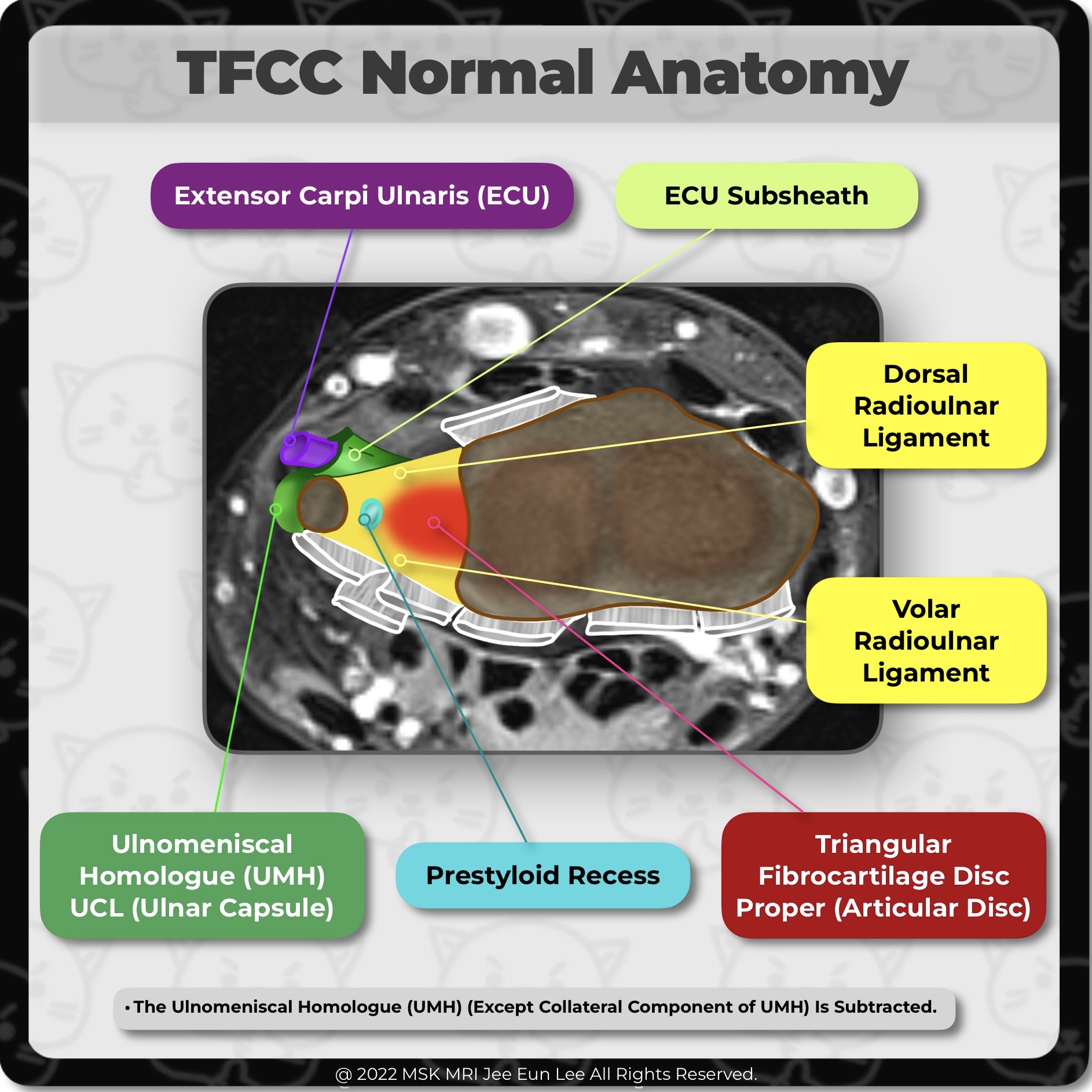
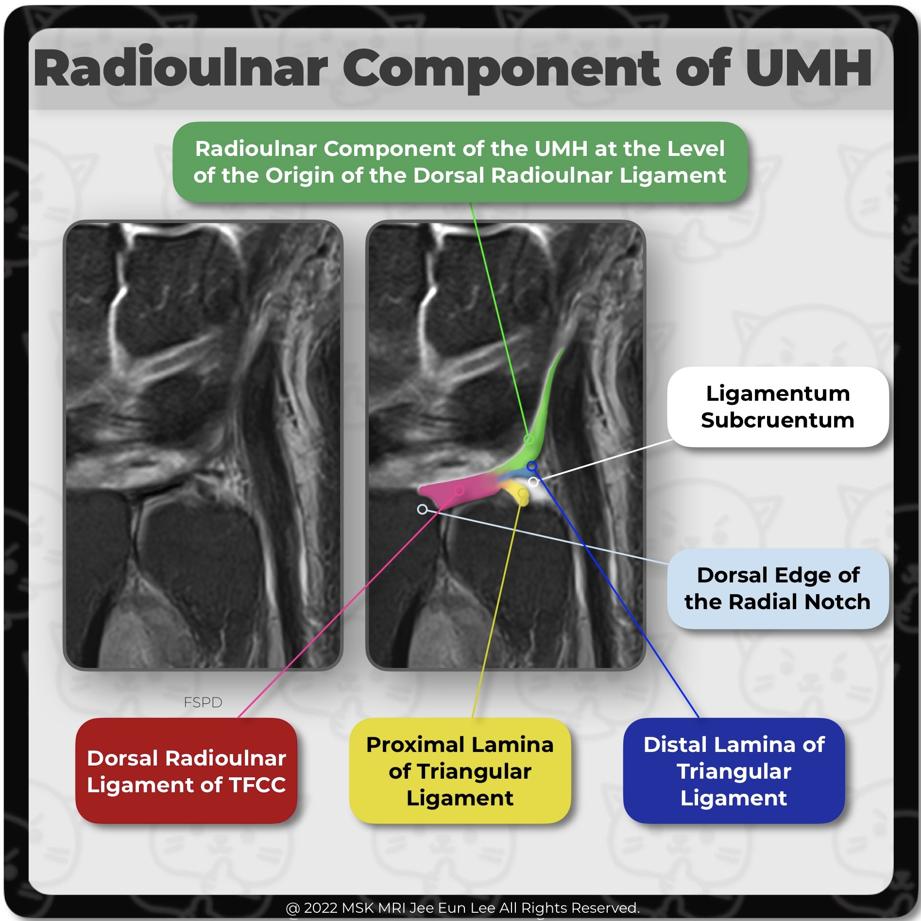
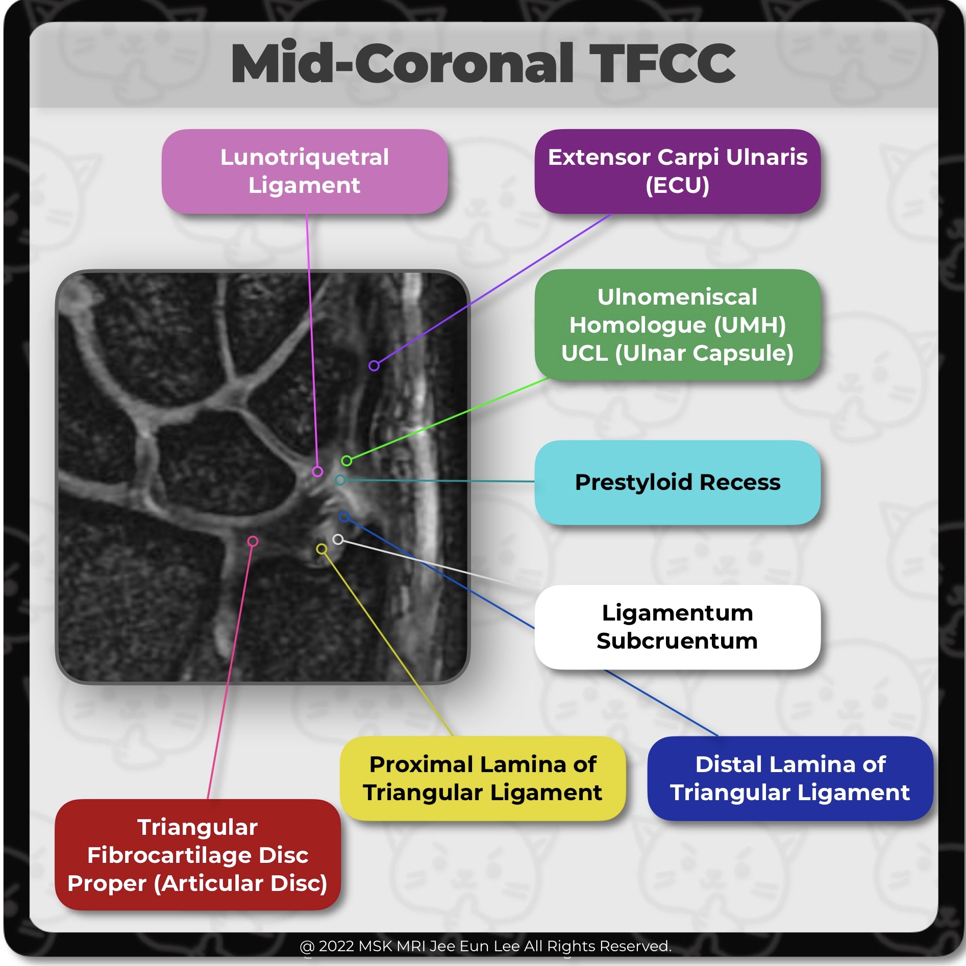
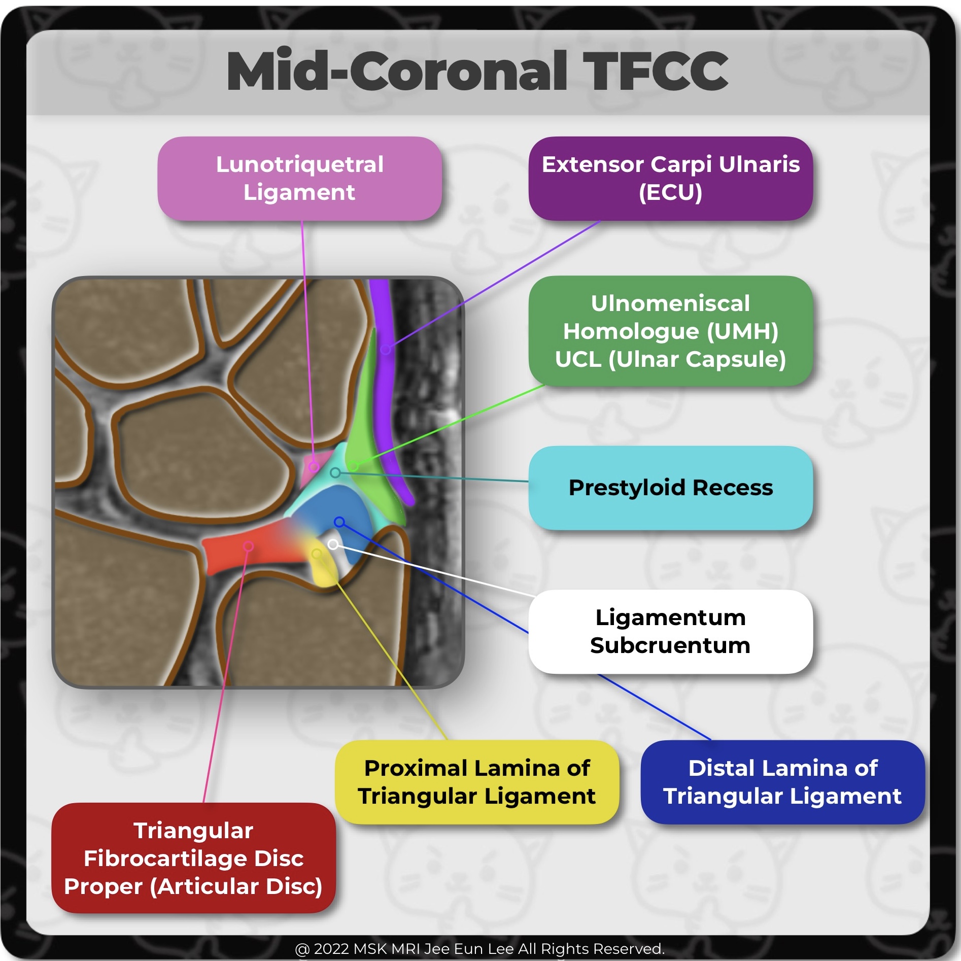
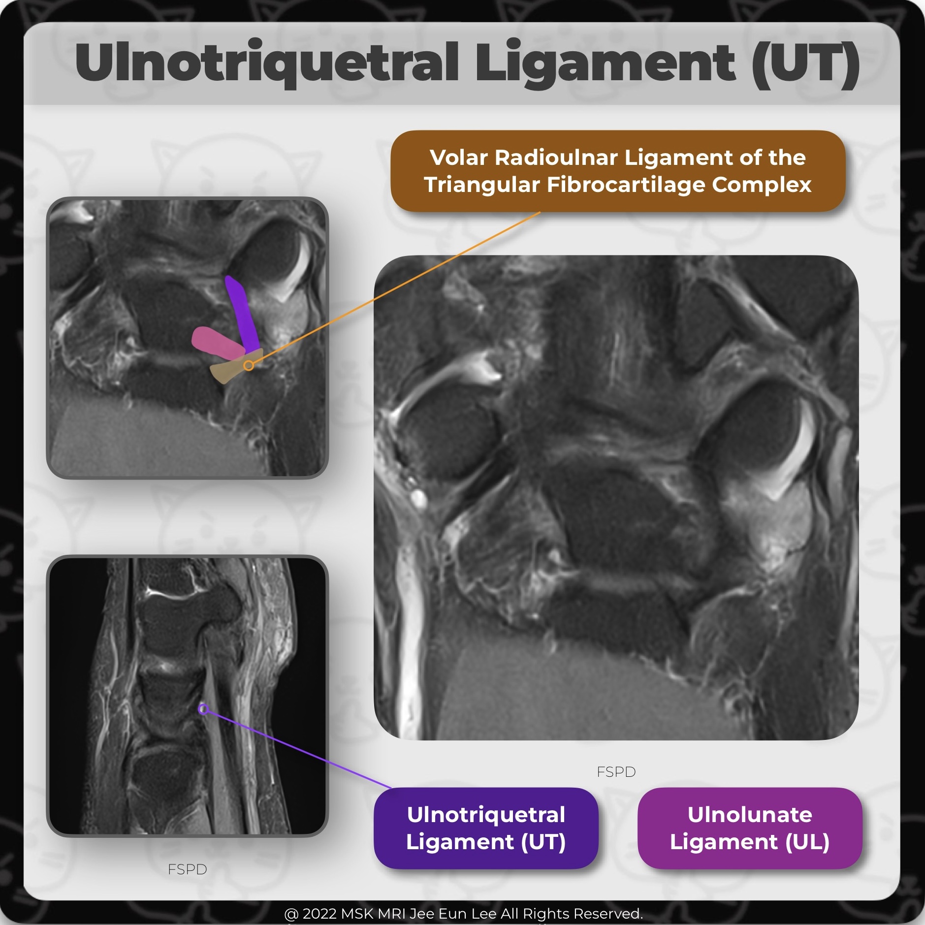
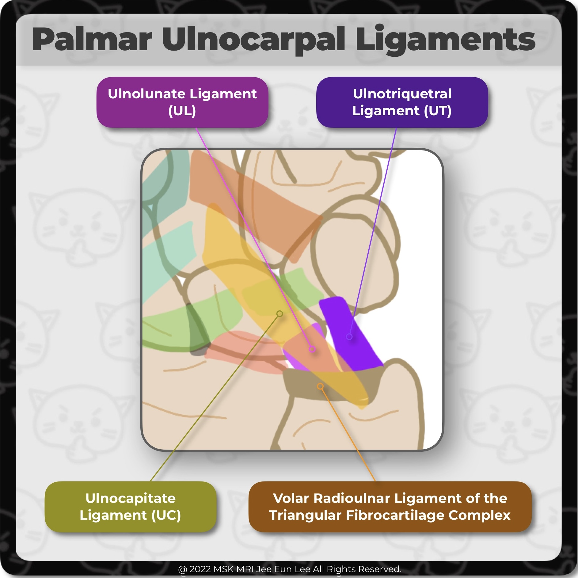
The most important structure of the triangular fibrocartilage complex (TFCC) is the TFC proper. The ulnar structures include the meniscus homologue (MH) and ulnar collateral ligament (ulnar capsule). The volar structures include the volar radioulnar ligament, ulnotriquetral and ulnolunate ligaments. The dorsal structures include the dorsal radioulnar ligament and extensor carpi ulnaris tendon sh.. 2022. 9. 11.
[Anatomy_19] Distal Insertion of the ulnomeniscal homologue, TFCC, wrist MRI The fibers at the main insertion into the ulnar aspect of the triquetrum are recognized between the triquetrum and the ECU tendon. Some fibers attach to the ulnar aspect of the pisiform (P), forming the dorsal capsular ligament of the pisotriquetral joint. The entire distal insertion was clearly separated from the volar extrinsic ligaments. #WristMRI, #WristAnatomy, #WristLigament, #WristInstabi.. 2022. 9. 4.
[Anatomy_18] Component of ulnomeniscal homologue, TFCC, wrist MRI (Styloid component) this component is recognized between the entrance to the prestyloid recess and the the extensor carpi ulnaris tendon (ECU). (Collateral component), UMH, the ECU tendon sheath, and the joint capsule (ulnar collateral ligament) is fused indistinguishably. (Distal insertion) The ulnar aspect of the triquetrum represented the main insertion site. #WristMRI, #WristAnatomy, #WristL.. 2022. 9. 4.
[Anatomy_17] Ulnomeniscal homologue, Radioulnar Ligament TFCC, Ulnotriquetral Ligament, Ulnotriquetral The ulnomeniscal homologue (UMH) is noted slightly ulnar to the origin of the dorsal radioulnar ligament and radioulnar component of the UMH. The radioulnar component of the UMH arose at the dorsal radioulnar ligament of the TFC complex. The distal insertion of the UMH is seen with its main body attaching to the triquetrum #WristMRI, #WristAnatomy, #WristLigament, #WristInstability, #TFCC, #Wris.. 2022. 9. 4.
[Anatomy_16] Radioulnar Component of ulnomeniscal homologue, TFCC, wrist MRI At the level of the origin of the dorsal radioulnar ligament, radioulnar component of the ulnomeniscal homologue (UMH) is noted. The radioulnar component of the UMH arose at the dorsal edge of the radial notch, together with the dorsal radioulnar ligament of the TFC complex. The broad ligamentous structure attaches to the dorsal edge of the radial notch and divides into the proximal and distal l.. 2022. 9. 4.
[Anatomy_15] Triangular fibrocartilage complex (TFCC), wrist MRI The volar and dorsal aspects of the attachment correspond to slip insertions of the radioulnar ligaments into bone. The central portion of the attachment appears as an insertion into the hyaline cartilage of the radius The triangular fibrocartilage or articular disc of the TFCC has a broad attachment to the hyaline cartilage of the sigmoid notch of the distal radius, whereas, the dorsal and vola.. 2022. 9. 4.
[Anatomy_14] Triangular fibrocartilage complex (TFCC), wrist MRI On the ulnar attachment, TFCC attaches onto the ulnar styloid process through the distal lamina and onto the fovea of distal ulna through the proximal lamina; they are separated by the ligamentum subcruentum. The most important structure of the TFCC is the triangular fibrocartilage (TFC) proper, a triangular-shaped piece of fibrocartilage. On the radial side, the triangular fibrocartilage or art.. 2022. 9. 4.
[Anatomy_13] Ulnotriquetral Ligament, Two Normal Perforation, prestyloid recess, pisotriquetral joint orifice The ulnotriquetral ligament (UT) often has 2 normal perforations: a proximal opening to the prestyloid recess, and a distal pisotriquetral joint orifice. (Left image) The prestyloid recess perforation is found just ulnar to the union between the proximal radioulnar ligament and the UT ligament. (Right image) The pisotriquetral orifice is anterior to the proximal articular surface of the triquetr.. 2022. 9. 2.
[Anatomy_12] Ulnotriquetral Ligament, TFCC, wrist MRI The ulnotriquetral ligament (UT) forms the anterior and ulnar part of the ulnocarpal joint capsule together with the ulnolunate ligament. UT shares a common origin and connects the volar radioulnar ligament and thus the triangular fibrocartilage complex to the triquetral bone. #WristMRI, #WristAnatomy, #WristLigament, #WristInstability, #TFCC, #Wrist MRI, #Wrist anatomy, #wrist ligament, #안산에이스병.. 2022. 9. 2.
[Anatomy_11] Palmar Ulnocarpal Ligaments, TFCC, wrist MRI IThe ulnocarpal ligament, or ulnocarpal ligamentous complex (UCLC), is anatomically divided into the ulnocapitate, ulnotriquetral, and ulnolunate ligaments. The palmar ulnocarpal ligaments are the superficial ulnocapitate and the deeper ulnolunate and ulnotriquetral ligaments. #WristMRI, #WristAnatomy, #WristLigament, #WristInstability, #TFCC, #Wrist MRI, #Wrist anatomy, #wrist ligament, #안산에이스병.. 2022. 9. 1.
[HYPERLINK] Wrist MRI checklist (SITE MAP) Wrist MRI checklist [Wrist_01] MRI check list, TFCC, triangular fibrocartilage complex, Palmer [Wrist_02] MRI Check liest, SLL, LTL, MRI check list, Scapholunate ligament, lunotriquetral ligament [Wrist_03] MRI checklist, scaphoid, lunate, Bone & joint, AVN, Kienbock’s Disease, ulnar side pain [Wrist_04] extensor, flexor tendons, tenosynovitis, tendinosis, intersection, MRI checklist [Wrist_05] .. 2022. 5. 10.
[HYPERLINK] 삼각 섬유연골 복합체 (Triangular FibroCartilage Complex, TFCC), Palmer classification, (SITE MAP) 삼각 섬유연골 복합체 (Triangular FibroCartilage Complex, TFCC)​ [TFCC_01] TFCC- 1(anatomy -1) / wrist MRI triangular fibrocartilage complex, TFCC ​ [TFCC_02] TFCC- 2(anatomy -2) / wrist MRI triangular fibrocartilage complex, TFCC [TFCC_03] TFCC- 3 (Palmer type 1A) / wrist MRI triangular fibrocartilage complex, TFCC [TFCC_04] TFCC- 4 (Palmer type 1B) / wrist MRI triangular fibrocartilage complex, TFCC [TF.. 2022. 5. 10.