WRIST MRI 27

[Anatomy_16] Radioulnar Component of ulnomeniscal homologue, TFCC, wrist MRI

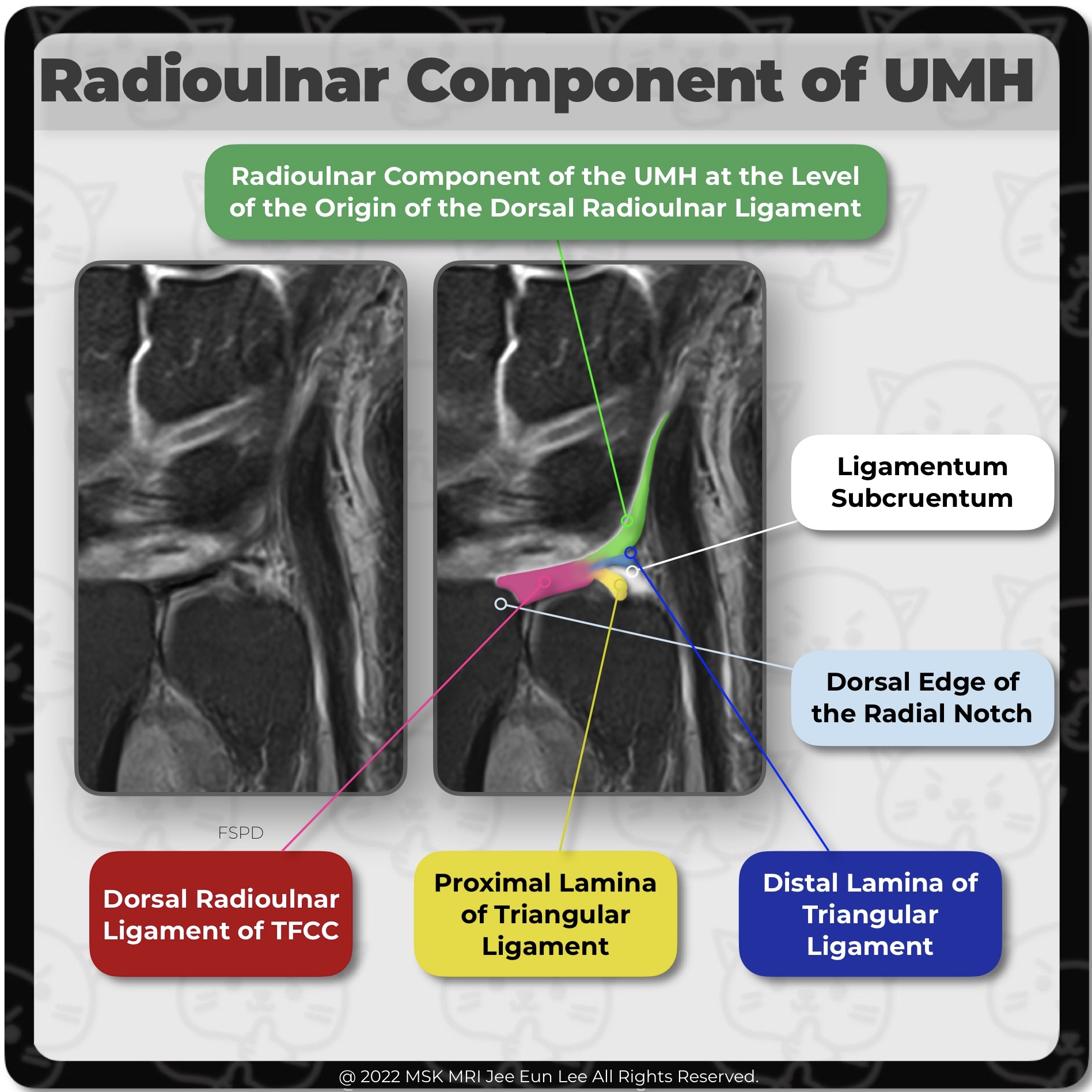
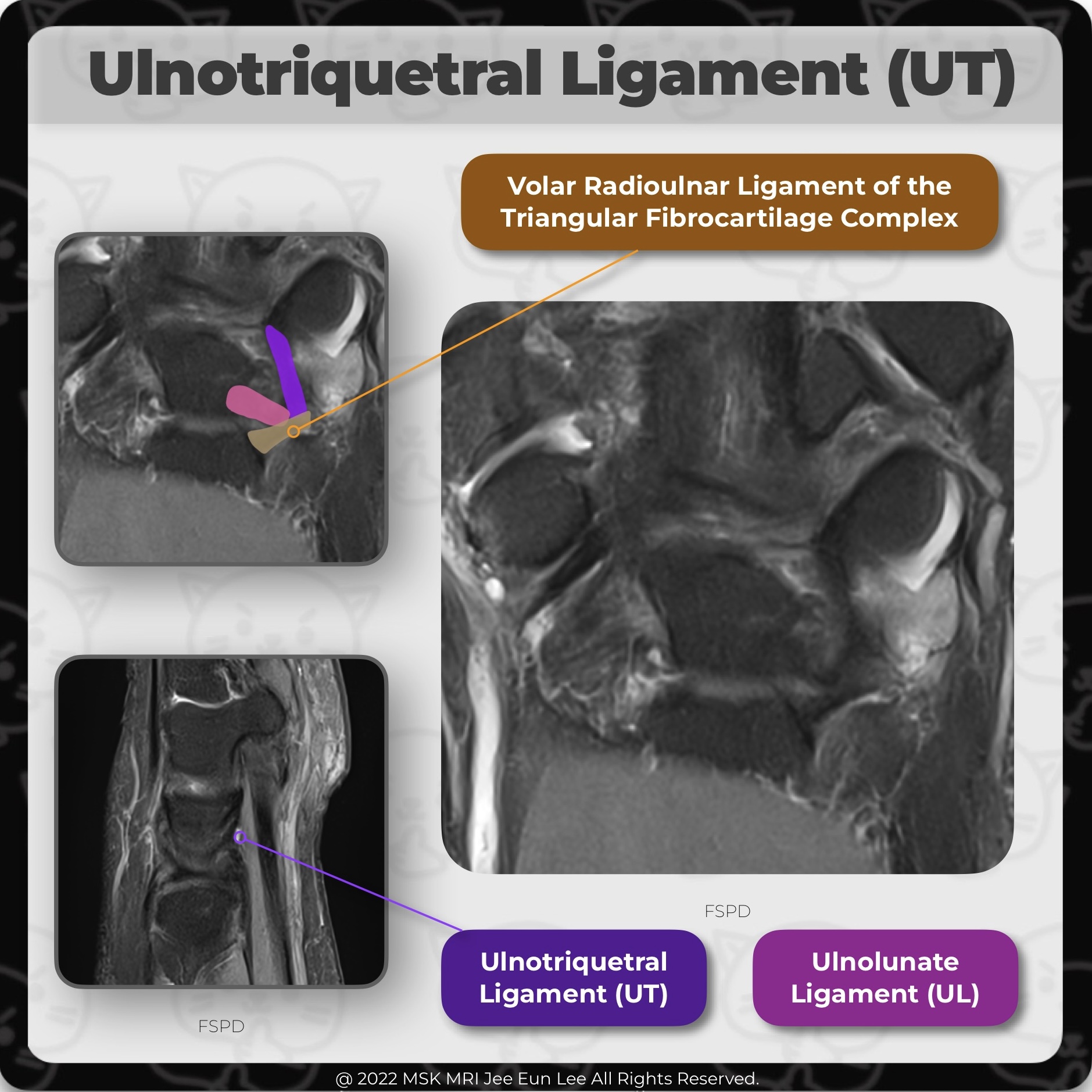
At the level of the origin of the dorsal radioulnar ligament, radioulnar component of the ulnomeniscal homologue (UMH) is noted. The radioulnar component of the UMH arose at the dorsal edge of the radial notch, together with the dorsal radioulnar ligament of the TFC complex. The broad ligamentous structure attaches to the dorsal edge of the radial notch and divides into the proximal and distal l..

[Anatomy_15] Triangular fibrocartilage complex (TFCC), wrist MRI

The volar and dorsal aspects of the attachment correspond to slip insertions of the radioulnar ligaments into bone. The central portion of the attachment appears as an insertion into the hyaline cartilage of the radius The triangular fibrocartilage or articular disc of the TFCC has a broad attachment to the hyaline cartilage of the sigmoid notch of the distal radius, whereas, the dorsal and vola..

[Anatomy_14] Triangular fibrocartilage complex (TFCC), wrist MRI

On the ulnar attachment, TFCC attaches onto the ulnar styloid process through the distal lamina and onto the fovea of distal ulna through the proximal lamina; they are separated by the ligamentum subcruentum. The most important structure of the TFCC is the triangular fibrocartilage (TFC) proper, a triangular-shaped piece of fibrocartilage. On the radial side, the triangular fibrocartilage or art..

[Anatomy_13] Ulnotriquetral Ligament, Two Normal Perforation, prestyloid recess, pisotriquetral joint orifice

The ulnotriquetral ligament (UT) often has 2 normal perforations: a proximal opening to the prestyloid recess, and a distal pisotriquetral joint orifice. (Left image) The prestyloid recess perforation is found just ulnar to the union between the proximal radioulnar ligament and the UT ligament. (Right image) The pisotriquetral orifice is anterior to the proximal articular surface of the triquetr..

[Anatomy_12] Ulnotriquetral Ligament, TFCC, wrist MRI

The ulnotriquetral ligament (UT) forms the anterior and ulnar part of the ulnocarpal joint capsule together with the ulnolunate ligament. UT shares a common origin and connects the volar radioulnar ligament and thus the triangular fibrocartilage complex to the triquetral bone. #WristMRI, #WristAnatomy, #WristLigament, #WristInstability, #TFCC, #Wrist MRI, #Wrist anatomy, #wrist ligament, #안산에이스병..

[Anatomy_11] Palmar Ulnocarpal Ligaments, TFCC, wrist MRI

IThe ulnocarpal ligament, or ulnocarpal ligamentous complex (UCLC), is anatomically divided into the ulnocapitate, ulnotriquetral, and ulnolunate ligaments. The palmar ulnocarpal ligaments are the superficial ulnocapitate and the deeper ulnolunate and ulnotriquetral ligaments. #WristMRI, #WristAnatomy, #WristLigament, #WristInstability, #TFCC, #Wrist MRI, #Wrist anatomy, #wrist ligament, #안산에이스병..

[Anatomy_10] Radioscapholunate Bundle, Scapholunate Ligament, wrist MRI

MRI shows the normal radioscapholunate bundle attaching to the junction of the volar and proximal segments of the SLL. Scaphoid (S) and lunate (L) bones. Note the low-signal ligamentous appearance of this neurovascular-containing mesocapsule. #WristMRI, #WristAnatomy, #WristLigament, #WristInstability, #Wrist MRI, #Wrist anatomy, #wrist ligament, #안산에이스병원,#Radioscapholunate Bundle, #Scapholunate..

[Anatomy_09] Short radiolunate ligament, Ulnolunate ligament, wrist MRI

The ulnolunate ligament(UL) is continuous with the short radiolunate ligament (SRL) and attaches to the palmar cortex of the lunate just as the short radiolunate ligament does. #WristMRI, #WristAnatomy, #WristLigament, #WristInstability, #Wrist MRI, #Wrist anatomy, #wrist ligament, #안산에이스병원,#short radiolunate ligament, #ulnolunate ligament, #ulnocarpal ligament, #ulnocarpal ligamentous complex,

[Anatomy_08] Scapholunate Ligament Tear, Radioscaphocapitate ligament, Long radiolunate ligament

FSPD image demonstrates full-thickness tear of the membranous portion (not shown) and volar band of the scapholunate ligament (SLL), and partial tear of dorsal band. Note the hyperintense signal and thickening of the radioscaphocapitate (RSC) ligament and long radiolunate ligament (LRL), consistent with grade I injury or sprain. LRL should not be mistaken for intact volar SLL. #WristMRI, #WristA..

[Anatomy_07] Scapholunate Ligament, Long radiolunate ligament, Radioscaphocapitate ligament

The lunate attachment is just dorsal to the long radiolunate ligament (LRL), (=radiolunotriquetral ligament (RLT)) attachment and immediately proximal to the cartilage surface. #WristMRI, #WristAnatomy, #WristLigament, #WristInstability, #Wrist MRI, #Wrist anatomy, #wrist ligament, #안산에이스병원, #Radioscaphocapitate ligament, #Long radiolunate ligament, #Scapholunate Ligament