본문 바로가기

lateral meniscus29

(Fig 1-A.12) Anatomy of popliteomeniscal fascicles 
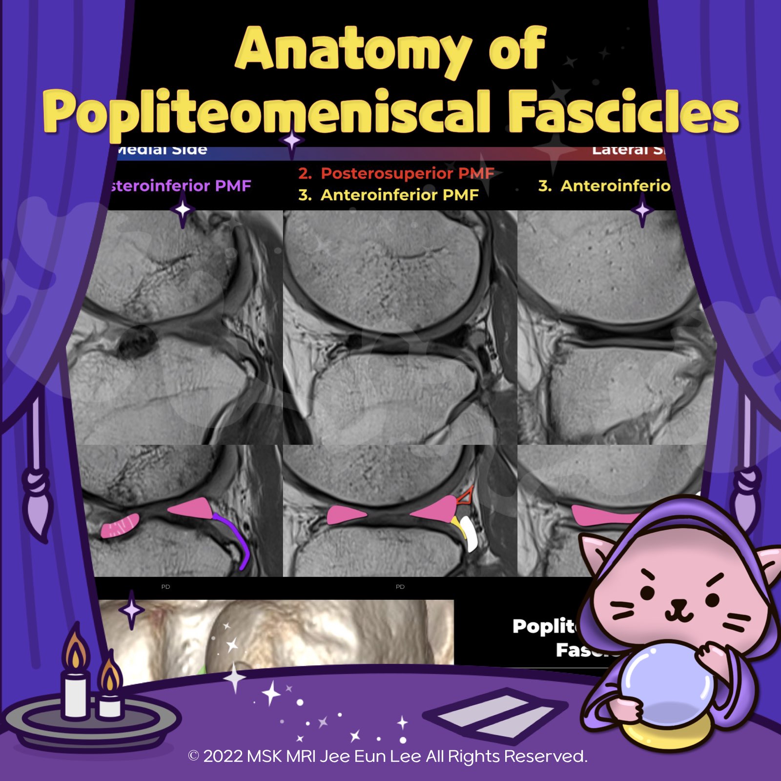
https://youtu.be/GYwJcbKhxbY Fascicle Location and Appearance Anteroinferior Popliteomeniscal Fascicle Visible on sagittal slices near the fibular head; extends posteroinferiorly from the lateral side of the lateral meniscus (LM) and merges with the popliteus tendon; conjoined attachment with the popliteofibular ligament at the fibula’s styloid process; variable appearance. Posterosuperior Popli.. 2024. 1. 10.
[Knee-01] Tear of anteroinferior popliteomeniscal fascicle, posterolateral corner injury https://youtu.be/he2-XqAyvaA * The popliteomeniscal fascicles arise from the periphery of the posterior horn of the lateral meniscus and form a posterolateral meniscocapsular extension, which creates the popliteus hiatus. * The fascicles not only form the hiatus, which allows the popliteus tendon an avenue to course intra-articularly and maintain the integrity of the joint but stabilize and cont.. 2022. 12. 21.
[Anatomy_30] Oblique Meniscomeniscal Ligament vs bucket handle tear -03 The medial OMML originates from the anterior horn of the medial meniscus and inserts onto the posterior horn of the lateral meniscus between the ACL and PCL. This structure should not be mistaken for a bucket handle meniscal tear A bucket-handle tear is a longitudinal tear with central migration of the inner “handle” fragment The correct diagnosis of an oblique meniscomeniscal ligament rather th.. 2021. 10. 2.
[Anatomy_29] Oblique Meniscomeniscal Ligament -02 The medial OMML originates from the anterior horn of the medial meniscus and extends through the intercondylar notch to insert onto the posterior horn of the lateral meniscus. This ligament passes between the ACL and PCL. This oblique meniscomeniscal ligament is kind of similar SI to meniscal tissue. The sagittal image near the midline demonstrates a linear low signal intensity structure atop th.. 2021. 10. 2.
[Anatomy_28] Oblique Meniscomeniscal Ligament -01 The medial and lateral oblique menisco-meniscal ligaments are rare and can mimic a tear if not recognized. The oblique meniscomeniscal ligament passes from the anterior horn of one meniscus to the posterior horn of the opposite meniscus. The oblique meniscomeniscal ligaments are named based on their anterior attachment site. The medial OMML originates from the anterior horn of the medial meniscu.. 2021. 10. 2.
[Anatomy_27] Anterior transverse meniscal ligament -02 Both horns of the lateral meniscus are triangular in shape and have very sharp points. In this case, we can this low-signal-intensity band like structure adjacent to the anterior horn of the lateral meniscus Both cases are normal finding, The transverse ligament has various diameters. The anterior root of MM inserts along the tibial eminence, and posterior fibers of the anterior horn attachment .. 2021. 9. 27.
[Anatomy_26] Anterior transverse meniscal ligament -01 The transverse ligament is a fibrous band that connects the anterior horns of the medial and lateral menisci. Axial and sagittal images demonstrate the course of the transverse ligament as a low-signal-intensity band traversing Hoffa’s infrapatellar fat pad. The transverse ligament can simulate an oblique tear adjacent to the anterior horn of the lateral meniscus © 2021 MSK MRI Jee Eun Lee All R.. 2021. 9. 27.
[Anatomy_24] Meniscofemoral ligament, Humphry ligament The meniscofemoral ligaments originate from the posterior horn of the lateral meniscus and insert onto the lateral aspect of the posterior medial femoral condyle, anterior to the PCL. notice, This cleft between the meniscus and meniscofemoral ligament may be mistaken for a meniscal tear. © 2021 MSK MRI Jee Eun Lee All Rights Reserved. #MSKMRI, #virtualMRI, #radiologist, #Knee_MRI, #MSKMRI_Knee, .. 2021. 9. 26.
[Anatomy_23] Meniscofemoral ligament, Wrisberg ligament, PseudoTear, Wrisberg Ri https://youtu.be/twVB-GMvcIg Coronal image demonstrates the posterior meniscofemoral ligament of Wrisberg (arrow) coursing obliquely downward from the medial femoral condyle to attach to the posterior horn of the lateral meniscus The meniscofemoral ligaments originate from the posterior horn of the lateral meniscus and insert onto the lateral aspect of the posterior medial femoral condyle, poste.. 2021. 9. 26.
[Shorts #05] Meniscofemoral ligament, Wrisberg ligament #shorts •This posteroblique illustration shows the relationship of meniscofemoral ligaments, lateral meniscus, and PCL. The meniscofemoral ligaments originate from the posterior horn of the lateral meniscus and insert onto the lateral aspect of the posterior medial femoral condyle, with the Humphry ligament anterior to the posterior cruciate ligament (PCL) and the Wrisberg ligament posteriorly Subset of.. 2021. 9. 26.
[Anatomy_22] Meniscofemoral ligament, Humphry, Wrisberg The major attachments of the posterior horn of the lateral meniscus are the popliteomeniscal fascicles and the meniscofemoral ligaments. This posteroblique illustration shows the relationship of meniscofemoral ligaments, lateral meniscus, and PCL. The meniscofemoral ligaments originate from the posterior horn of the lateral meniscus and insert onto the lateral aspect of the posterior medial femo.. 2021. 9. 26.
[Shorts #03] Meniscofemoral ligament, Humphry, Wrisberg #shorts •This figure illustrates the relationship of meniscofemoral ligaments, lateral meniscus, and PCL. The meniscofemoral ligaments originate from the posterior horn of the lateral meniscus and insert onto the lateral aspect of the posterior medial femoral condyle, with the Humphry ligament anterior to the posterior cruciate ligament (PCL) and the Wrisberg ligament posteriorly Occasionally the menisc.. 2021. 9. 25.
[Anatomy_21] Posteroinferior popliteomeniscal fascicle The popliteomeniscal fascicles consist of the anteroinferior(strongest), posterosuperior, and posteroinferior. posteroinferior The posteroinferior popliteomeniscal fascicle (arrow) is the least consistently visualized of the fascicles The posteroinferior fascicle is more medially located, and extends from a medial aponeurotic extension of the popliteus tendon to attach to the lateral meniscus ne.. 2021. 9. 25.
[Anatomy_20] Posterosuperior popliteomeniscal fascicle The popliteomeniscal fascicles arise from the periphery of the posterior horn of the lateral meniscus and form a posterolateral meniscocapsular extension, which creates the popliteus hiatus. The popliteal hiatus shows relatively symmetric posterosuperior and anteroinferior fascicles. More medial section of the hiatus shows the normal deficiency of the anteroinferior popliteomeniscal fascicle. le.. 2021. 9. 25.
[Anatomy_18] Anteroinferior popliteomeniscal fascicle 01 The anteroinferior popliteomeniscal fascicle (yellow arrow) is seen to course from the inferior aspect of the lateral meniscus posteroinferiorly to blend with fibers of the popliteofibular ligament This fascicle is typically seen on sagittal slices where the fibular head is also visible. In another patient, a component of the anteroinferior popliteomeniscal fascicle which courses directly to the.. 2021. 9. 25.
[Anatomy_17] Tear of posterosuperior Popliteomeniscal fascicle The posterosuperior popliteomeniscal fascicle forms the roof of the popliteus hiatus, extending from the posterosuperior margin of the lateral meniscus to the posterior capsule. The anteroinferior popliteomeniscal fascicle is a coronary ligament, which forms the floor of the popliteus hiatus. As the popliteus tendon passes laterally, it passes beneath the posterosuperior fascicle and above the a.. 2021. 9. 25.
[Anatomy_16] Overview of the popliteomeniscal fascicles The LM has a loose attachment to the joint capsule and to the femur and tibia by means of the meniscal ligaments and is not anchored to the lateral collateral ligament (LCL). Posterior oblique drawing demonstrates posterolateral restraints of the lateral meniscus including the posterosuperior popliteomeniscal fascicle (PSF), anteroinferior popliteomeniscal fascicle (AIF), meniscofemoral ligament.. 2021. 9. 24.
[Anatomy_15] Lateral Meniscus Posterior Root - 03 PRLM has two attachment sites of the intertubercular region and the posterior slope of the lateral tubercle. Contiguous fat-suppressed PD coronal images with a posteroanterior direction demonstrate small, dark signal foci (white arrows) traversing the intertubercular crest and terminating on the medial tubercle, near the tibial attachment of the ACL. These green-yellow arrows show an intercondyl.. 2021. 9. 24.
[Anatomy_14] Lateral Meniscus Posterior Root - 02 PRLM has two attachment sites of intertubercular region and posterior slope of lateral tubercle. This image shows an intercondylar crest insertion component among the two attachment sites. The PRLM inserts mainly in the intertubercular area. It is delineated on MRI as a dark signal line parallel to the tibial plateau on mid-sagittal images with an oblique course and longer length than other root.. 2021. 9. 24.
[Anatomy_13] Lateral Meniscus Posterior Root - 01 The PRLM(lateral meniscus posterior root) provides secondary stability to the anterior cruciate ligament during pivot shift loading. The PRLM has unique features, including its course and configuration. An accurate anatomic understanding of the PRLM is clinically important because this region is difficult to fully visualize arthroscopically. PRLM has two attachment sites of intertubercular regio.. 2021. 9. 24.
[Anatomy_12] Normal Fissured LM Anterior Root The anterior root of lateral meniscus is normally directed obliquely upward on sagittal images and frequently contains increased internal signal intensity. In this location, the increased signal intensity is sometimes referred to as “speckled.” The anterior root of the lateral meniscus differs from the anterior root of the medial meniscus because it often has a prominent fissured appearance simi.. 2021. 9. 24.