Click the link to purchase on Amazon 🎉📚
==============================================
🎥 Check Out All Videos at Once! 📺
👉 Visit Visualizing MSK Blog to explore a wide range of videos! 🩻
https://visualizingmsk.blogspot.com/?view=magazine
📚 You can also find them on MSK MRI Blog and Naver Blog! 📖
https://www.instagram.com/msk_mri/
Click now to stay updated with the latest content! 🔍✨
==============================================
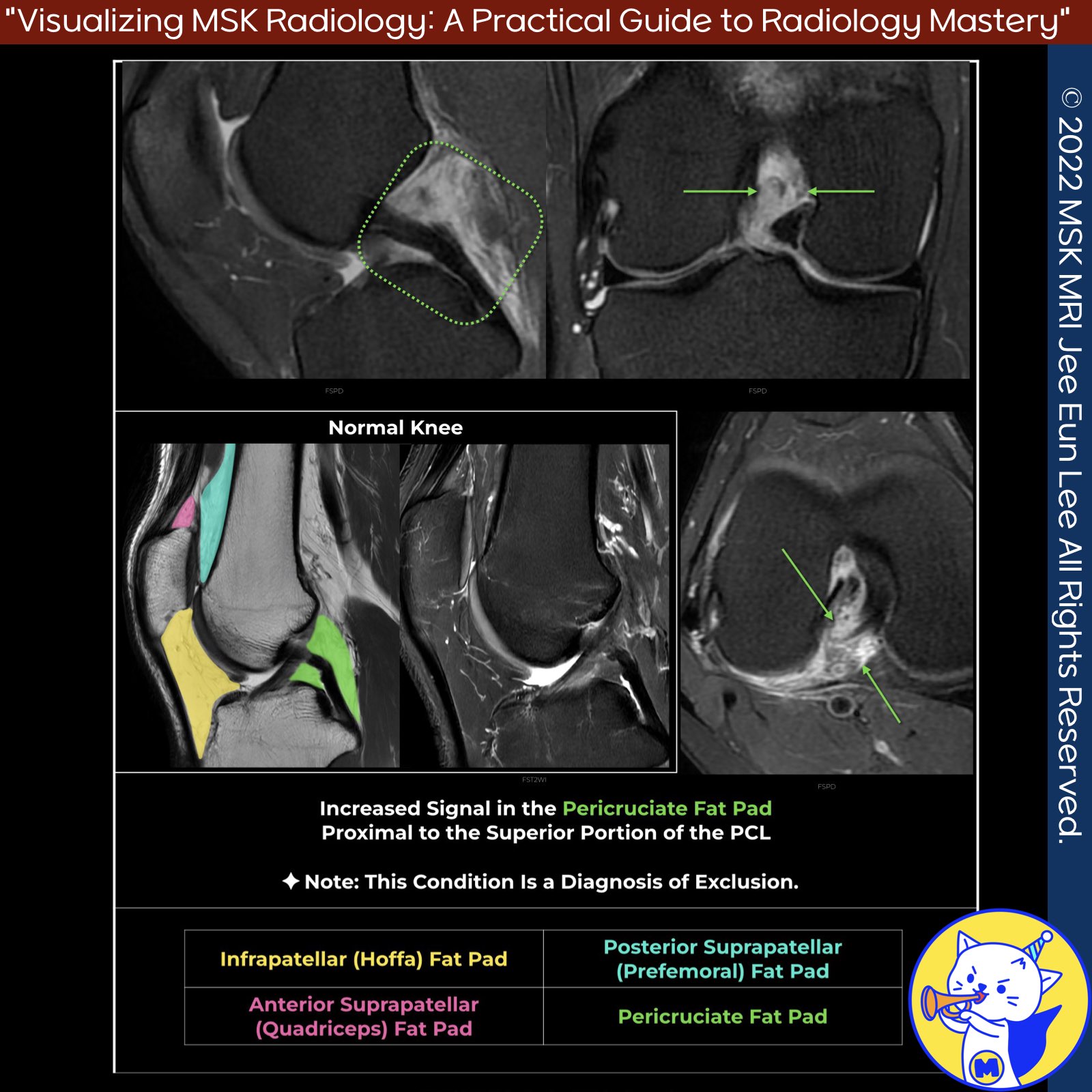
📌Peri-cruciate Fat Pad Inflammation
- Peri-cruciate fat pad inflammation commonly presents as non-specific posterior knee pain, particularly in young individuals with high physical activity levels.
- This condition is often associated with the impingement of the fat pad during knee flexion and is considered an exclusion diagnosis.
✅MRI Findings:
- Poorly defined edema in the peri-cruciate fat pad on fluid-sensitive fat-suppressed sequences.
- Best appreciated on sagittal and axial images.
- The edematous fat pad enhances following contrast administration.
📌Adhesive Capsulitis of the Knee
- Adhesive capsulitis of the knee involves a combination of synovial inflammation and capsular fibrosis.
- The disease progresses from synovial inflammation to fibroblastic infiltration, leading to adhesion and retraction of the capsule.
- Typically, pain presents first, followed by a progressive loss of motion; the pain then subsides, and motion may or may not be slowly restored .
✅ MRI Findings:
- High signal intensity on fluid-sensitive images.
- Contrast enhancement of the pericapsular tissues, especially around the anterior cruciate ligament, popliteal tendon insertion, and posteromedial and lateral capsule.
- Quadriceps fat pad involvement, with signal abnormalities most likely representing edema.
- This overlap suggests that isolated suprapatellar fat pad signal alteration may be a forme fruste ( incomplete phenotypic expression of a condition, such that it does not meet the usual diagnostic criteria.) of adhesive capsulitis.
References
- Skeletal Radiology (2020) 49:823–836
- Magn Reson Imaging Clin N Am 22 (2014) 725–741
"Visualizing MSK Radiology: A Practical Guide to Radiology Mastery"
© 2022 MSK MRI Jee Eun Lee All Rights Reserved.
No unauthorized reproduction, redistribution, or use for AI training.
#PeriCruciateFatPadInflammation, #PosteriorKneePain, #YoungAthletes, #KneeFlexion, #DiagnosisOfExclusion, #MRI, #Oedema, #AdhesiveCapsulitis, #SynovialInflammation, #CapsularFibrosis
'✅ Knee MRI Mastery > Chap 4BCD. Anterior knee' 카테고리의 다른 글
| (Fig 4-C.09) Medial Plica Syndrome (0) | 2024.06.19 |
|---|---|
| (Fig 4-C.08) Anatomy of Patellar Plicae (0) | 2024.06.18 |
| (Fig 4-C.06) Quadriceps Fat Pad Edema (0) | 2024.06.18 |
| (Fig 4-C.05) Pre-femoral Fat Pad (0) | 2024.06.17 |
| (Fig 4-C.04) Postoperative Changes of Hoffa’s Fat Pad (0) | 2024.06.17 |


